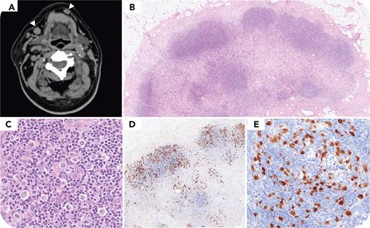
A 71-year-old man presented with a 2-month history of fever of unknown origin. No lymphadenopathy, except for mild submandibular lymph node enlargement, was detected on computed tomography (panel A, arrowheads) and 18F-fluorodeoxyglucose positron emission tomography. Biopsies of the submandibular lymph node revealed lymphoid nodules composed of scattered large cells with irregular nuclei surrounded by fibrotic tissue (panel B: hematoxylin and eosin stain, original magnification ×40; panel C: hematoxylin and eosin stain, original magnification ×400). Immunohistochemical analysis revealed large cells positive for CD30 (panel D: original magnification ×40); anaplastic lymphoma kinase (ALK), which showed cytoplasmic, nuclear, and nucleolar staining patterns (panel E: original magnification ×400); CD4; epithelial membrane antigen; granzyme B; T-cell intracellular antigen 1; and programmed death ligand 1. However, the cells were negative for CD20, CD3, CD15, PAX5, CD5, CD7, CD8, GATA3, and Epstein-Barr virus–encoded small RNA on in situ hybridization. Ki-67 showed 100% positivity. Fluorescence in situ hybridization showed an ALK split signal. The patient was diagnosed with ALK-positive anaplastic large cell lymphoma (ALK+ ALCL) with a Hodgkin-like pattern and was treated with brentuximab vedotin, cyclophosphamide, doxorubicin, and prednisone. His fever resolved after initiation of treatment.
ALK+ ALCL exhibits a Hodgkin-like growth pattern resembling nodular sclerosis classical Hodgkin lymphoma in 2.6% of cases. When tumor cells are PAX5 negative, despite nodular sclerosis and a classical Hodgkin lymphoma–like histopathology, ALK immunohistochemistry should be performed based on the assumption of an ALK+ ALCL, Hodgkin-like pattern.
For additional images, visit the ASH Image Bank, a reference and teaching tool that is continually updated with new atlas and case study images. For more information, visit https://imagebank.hematology.org.


This feature is available to Subscribers Only
Sign In or Create an Account Close Modal