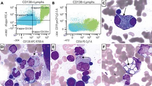
A 66-year-old man with a 15-year history of immunoglobulin G κ multiple myeloma and status post–autologous stem cell transplant and multiple lines of therapy (most recently, daratumumab, cyclophosphamide, and dexamethasone as a bridge to CAR-T therapy) presented for a bone marrow biopsy due to evidence of disease progression. Imaging showed hepatic lesions, and M-protein concentration was 1.78 g/dL (increased from 1.06 g/dL 1 month earlier). Flow cytometry of the aspirate showed involvement by a monotypic plasma cell population that expressed CD138, CD27, CD56, and κ immunoglobulin light chain (panels A-B; lime green: plasma cells). A differential count of the bone marrow aspirate showed 13% plasma cells, some with binucleation or nucleoli. A subset of cells showed coarse azurophilic granules (panels C-E [arrows]; Wright stain, 100× objective), morphologically consistent with Snapper-Schneid bodies. Some cells additionally showed vacuolization (panel F [arrow]; Wright stain, 100× objective).
This case displays Snapper-Schneid bodies, an uncommon cytologic feature most commonly seen in neoplastic plasma cells. Originally described in Blood in 1947 in patients who had received diamidine treatment, these granules of lysosomal origin have also been seen in patients receiving modern therapies and in rare cases of reactive plasmacytosis.
For additional images, visit the ASH Image Bank, a reference and teaching tool that is continually updated with new atlas and case study images. For more information, visit https://imagebank.hematology.org.


This feature is available to Subscribers Only
Sign In or Create an Account Close Modal