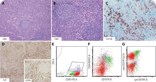
A 7-month-old boy with a history of biliary atresia presented with abdominal distension. Laboratory data showed anemia, thrombocytopenia, hypoalbuminemia, hypergammaglobulinemia, and elevated interleukin-6 and erythrocyte sedimentation rate. Imaging showed extensive abdominal and retroperitoneal lymphadenopathy. Lymph node biopsy demonstrated features of Castleman disease, mixed variant (panel A-B, hematoxylin and eosin [H&E] stain, 4× objective, 20× objective, respectively). Interfollicular areas showed prominent vascularity with increased CD138+ plasma cells (panel C, CD138 stain, 10× objective) and frequent terminal deoxynucleotidyltransferase–positive (TdT+) CD99+ T-cells (panel D, TdT stain, 4× objective; inset, CD99 stain, 10× objective) lacking overt cytologic atypia. Flow cytometry identified a discrete population of immature T cells (green population) expressing dim CD45 (panel E [SSC-A, side scatter; FITC-A, fluorescein isothiocyanate]), CD34 (panel F), CD10 (panel F [PE-A, phycoerythrin]), TdT (panel G [APC-A, allophycocyanin]), CD1a, CD2, cytoplasmic CD3 (panel G), CD5, and CD7 and lacking surface CD3, CD4, CD8, and surface T-cell receptor (TCR). TCRβ and TCRγ polymerase chain reaction studies were negative for clonal rearrangement. A human herpesvirus 8 (HHV-8) latency-associated nuclear antigen immunostain was negative. The clinicopathologic findings were consistent with idiopathic multicentric Castleman disease (iMCD), HHV-8−, with a modest proliferation of immature T cells.
iMCD is extremely uncommon in children, and whether its underlying pathophysiology differs from that observed in adults remains uncertain. Clinically indolent T-lymphoblastic proliferations (iT-LBP) can be seen in Castleman disease and may show features mimicking T-cell malignancies. However, iT-LBPs lack significant morphologic atypia, aberrant immunophenotype, and monoclonality and do not involve bone marrow or mediastinum.
For additional images, visit the ASH Image Bank, a reference and teaching tool that is continually updated with new atlas and case study images. For more information, visit https://imagebank.hematology.org.


This feature is available to Subscribers Only
Sign In or Create an Account Close Modal