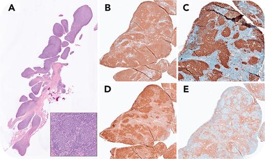
A 79-year-old woman with a prior follicular lymphoma (FL) diagnosed in 2007 underwent knee replacement for long-standing rheumatoid arthritis (RA). At the time of surgery the patient was in remission, with negative imaging studies. The synovial lining showed striking involvement by FL (panel A, original magnification ×1.25, hematoxylin and eosin stain). Expanded villous structures were infiltrated by abnormal follicles composed of centrocytes (panel A inset, original magnification ×40). The atypical follicles were strongly positive for CD20 (panel B, original magnification ×5, immunohistochemical staining), CD10 (panel C, original magnification ×5, immunohistochemical staining), and BCL2 (panel D, original magnification ×5, immunohistochemical staining) with surrounding interfollicular CD3+ T cells (panel E, original magnification ×5, immunohistochemical staining). The neoplastic follicles contained CD21+ follicular dendritic cell meshworks (not shown). The bone marrow space was negative for lymphoma.
Synovial tissues in RA can show prominent lymphoid aggregates with reactive germinal centers, seen in ∼25% of patients with well-established disease. The reactive follicles express activation-induced cytidine deaminase. FL B cells show homing to germinal centers throughout the lymphoid system, but involvement of the synovium in autoimmune conditions has not been well described. The synovial involvement in this patient was clinically silent, with the symptoms related to long-standing joint damage. Local chronic inflammatory stimulation likely played a role in the unique pattern of dissemination seen in this case.
For additional images, visit the ASH Image Bank, a reference and teaching tool that is continually updated with new atlas and case study images. For more information, visit https://imagebank.hematology.org.


This feature is available to Subscribers Only
Sign In or Create an Account Close Modal