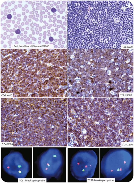
A 68-year-old man presented with fatigue and weight loss, and imaging showed bilateral neck and supraclavicular lymphadenopathy, enlargement of bilateral palatine tonsils and lingual tonsils, and bilateral axillary, retroperitoneal, external iliac, and inguinal lymphadenopathy. Complete blood count showed mild leukocytosis, and peripheral smear showed atypical lymphoid cells with occasional cells having nucleoli and cytoplasmic blebs (Giemsa stain; 100× objective). Lymph node biopsy showed an effaced architecture composed of small to medium-sized lymphoid cells diffusely positive for CD3, CD4, CD8, and TCL1 (all images using 60× objective). Lymphoid cells also expressed CD5 and CD7 and were negative for CD34, terminal deoxynucleotidyltransferase, and CD1a (images not shown). Bone marrow biopsy showed involvement by the abnormal T-cell population with a complex karyotype but without inv14 or t(14;14) translocation. Using break-apart probes, fluorescence in situ hybridization analysis identified TCL1 and TCRB rearrangements, and TCRA/D genes on chromosome 14 were intact (data not shown). The diagnosis of T-cell prolymphocytic leukemia/lymphoma (T-PLL) was made.
T-PLL is characterized by chromosomal rearrangement of inv14(q11q32) or t(14;14)(q11;q23) juxtaposing TCL1A gene next to TCRA/D. In 5% of the cases, t(X;14)(q28;q11.2) results in MTCP1::TCRA/D rearrangement. Translocation of the TCL1A gene with TCRB has been reported in rare situations.
For additional images, visit the ASH Image Bank, a reference and teaching tool that is continually updated with new atlas and case study images. For more information, visit https://imagebank.hematology.org.


This feature is available to Subscribers Only
Sign In or Create an Account Close Modal