In this issue of Blood, Mohammed et al report a complete atomic-resolution structure of factor V (FV) short, an enigmatic splicing variant of coagulation FV associated with bleeding.1 This structure, which includes the whole FV short B domain, provides valuable clues to understanding the unique functional properties of FV short.
FV (reviewed by Dahlbäck2) is a 330-kDa single-chain glycoprotein comprising multiple domains (A1-A2-B-A3-C1-C2). A high-affinity electrostatic interaction between a basic region (BR) and an acidic region (AR) within the large B domain (836 amino acids) maintains FV in the inactive state. Limited proteolysis at R709, R1018, and R1545 by factor Xa (FXa) and/or thrombin removes the B domain and converts FV to its active form (FVa), which acts as an essential cofactor of FXa in the prothrombinase complex. FVa activity is regulated by proteolytic cleavage at R306 and R506 by activated protein C (APC) and its cofactor protein S (PS).
The elucidation of the etiology of the East Texas bleeding disorder revealed the existence of a minor splicing isoform of FV, known as FV short, that circulates in plasma at a concentration of ∼0.2 nM.3 FV short is characterized by a large deletion (702 amino acids) within the B domain, which removes the BR and leaves the AR unmatched. This makes FV short constitutively active as a prothrombinase cofactor,4 suggesting it has a procoagulant function. However, in plasma FV short forms a trimolecular complex with tissue factor pathway inhibitor (TFPIα) and PS,2 which effectively suppresses the prothrombinase activity of FV short through a tight interaction between a BR in the C-terminus of TFPIα and the exposed AR of FV short.4 Bound TFPIα also protects FV short from cleavage at R1545, thereby preventing the separation of the AR (and hence TFPIα) from FV short.5 In turn, FV short maintains TFPIα in the circulation3 and, together with PS, potently enhances the inhibition of FXa by TFPIα.2 Therefore, the physiological interaction with TFPIα and PS converts FV short from a potentially procoagulant to an anticoagulant cofactor, explaining the bleeding tendency associated with elevated levels of the FV short/TFPIα/PS complex.3 Yet the exact role of FV short in the regulation of coagulation remains elusive.
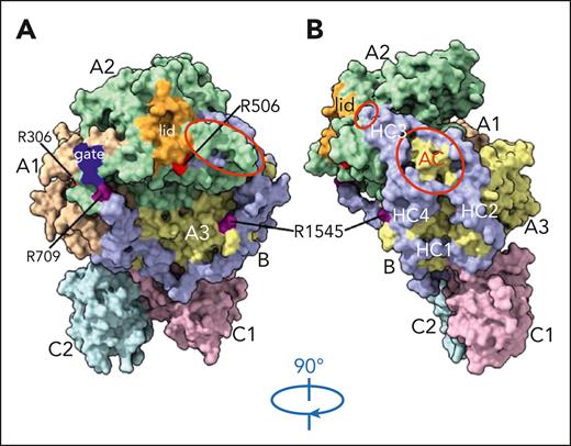
The structure of FV short (see figure) offers a mechanistic framework to interpret this body of experimental observations and to start unravelling the complex physiology of FV short. A major asset of this structure is the resolution of the entire FV short B domain, including the AR, which contains the main interaction site for TFPIα, and the pre-AR region, which is required for synergy with PS in the TFPIα-mediated inhibition of FXa.2
Structure of FV short. The structure, shown in front (A) and side (B) views, includes all residues of the native FV short protein (except 1 B domain residue encoded by the codon that spans the FV short splicing junction). The A1 (wheat), A2 (pale green), and A3 (pale yellow) domains are arranged in a triangular fashion on top of the phospholipid binding C1 (light pink) and C2 (pale cyan) domains. The B domain (light blue) spans from R709 to R1545 and wraps around the A domains. The thrombin-cleavage sites at R709 and R1545, as well as the partially exposed APC-cleavage sites at R306 and R506, are shown. The “gate” and “lid” regions of the A2 domain are highlighted in blue and orange, respectively. The exposed acidic segment of the A2 domain and the portions of the B (and A3) domains contributing to the negatively charged surface that may serve as binding site for the protease domain of FXa and the basic region of TFPIα are encircled in red. The approximate locations of the 4 hydrophobic clusters (HCs) surrounding the acidic cluster (AC) are marked in white. HC2 contains the PLVIVG hydrophobic patch required for synergy with PS in the TFPIα-mediated inhibition of FXa. Adapted from Figure 2 in the article by Mohammed et al that begins on page 3215.
Structure of FV short. The structure, shown in front (A) and side (B) views, includes all residues of the native FV short protein (except 1 B domain residue encoded by the codon that spans the FV short splicing junction). The A1 (wheat), A2 (pale green), and A3 (pale yellow) domains are arranged in a triangular fashion on top of the phospholipid binding C1 (light pink) and C2 (pale cyan) domains. The B domain (light blue) spans from R709 to R1545 and wraps around the A domains. The thrombin-cleavage sites at R709 and R1545, as well as the partially exposed APC-cleavage sites at R306 and R506, are shown. The “gate” and “lid” regions of the A2 domain are highlighted in blue and orange, respectively. The exposed acidic segment of the A2 domain and the portions of the B (and A3) domains contributing to the negatively charged surface that may serve as binding site for the protease domain of FXa and the basic region of TFPIα are encircled in red. The approximate locations of the 4 hydrophobic clusters (HCs) surrounding the acidic cluster (AC) are marked in white. HC2 contains the PLVIVG hydrophobic patch required for synergy with PS in the TFPIα-mediated inhibition of FXa. Adapted from Figure 2 in the article by Mohammed et al that begins on page 3215.
The overall arrangement of the A and C domains of FV short appears similar to that of FV,6 whereas the B domain wraps around the A domains (see figure). The authors point out specific differences in the “gate” and “lid” regions of the A2 domain that may account for the constitutive prothrombinase activity of FV short, and show that both FXa and prothrombin can be docked onto the FV short structure without significant hindrance by the B domain. Moreover, they identify an extended negatively charged surface, arising from the juxtaposition of acidic residues of the B domain (AR) and the C-terminal portion of the A2 domain, as the putative interaction site for the protease domain of FXa as well as the BR region of TFPIα (see figure). FXa binding would elicit the prothrombinase activity of FV short, whereas TFPIα binding would inhibit this activity. This regulatory mechanism, based on competition between FXa and TFPIα for the same binding site, could apply to other FV species that lack the BR but retain the AR, such as partially activated FV and platelet FV.4,7 Moreover, the interaction between the BR of TFPIα and the AR of FV short may serve as a model for the presently unresolved6 intramolecular interaction between the BR and AR of FV.
Interestingly, the FV short structure also suggests that TFPIα binding to FV short would mask the thrombin-cleavage site at R1545 and the APC-cleavage site at R506 (see figure). Both predictions are consistent with experimental evidence.5,8 In addition, the involvement of the R506 residue is supported by the observation that the R506Q mutation (FVLeiden) interferes with prothrombinase inhibition by TFPIα, which contributes to the risk of venous thrombosis associated with FVLeiden.9
Another remarkable feature of the FV short structure is the striking constellation of potential interaction sites created by the B domain on the surface of the A3 and A2 domains, where the pre-AR and AR regions define an acidic cluster surrounded by 4 hydrophobic clusters (see figure panel B). Notably, the second hydrophobic cluster contains a hydrophobic patch (PLVIVG, residues 1481-1486 of the pre-AR region) that in mutagenesis studies has proved essential for the cooperative assembly of the FV short/TFPIα/PS complex and for the synergy between FV short and PS as cofactors of TFPIα in the inhibition of FXa.2 This epitope has been proposed to interact with a stretch of hydrophobic residues (LIKT) that precede the BR in the C-terminus of TFPIα, thereby strengthening the FV short/TFPIα interaction and creating a high-affinity binding site for PS.2 This hypothesis and the role of the other hydrophobic clusters await experimental testing, also in the light of parallel PS mutagenesis studies.10
In summary, the FV short structure represents a valuable tool for structure/function analyses of FV short (and other molecular forms of FV) and paves the way to detailed understanding of the FV short/TFPIα/PS complex and its interaction with FXa at the onset of coagulation. TFPIα, which binds all other components directly, is likely to stabilize the quaternary complex and would need to dissociate to unleash the procoagulant activities of FXa and FV short and allow coagulation to take off. A possible player in this process could be platelet polyphosphate, which has been shown to abrogate prothrombinase inhibition by TFPIα in purified systems.7 The physiological regulation of the TFPIα/PS/FV short/FXa complex deserves further investigation and may have potential as a therapeutic target.
Finally, the FV short structure and the accumulating functional insights will hopefully boost the development of a much-needed FV short assay, which would make it possible to document the interindividual variation in plasma FV short levels and to test its association with clinical end points.
Conflict-of-interest disclosure: The author declares no competing financial interests.


This feature is available to Subscribers Only
Sign In or Create an Account Close Modal