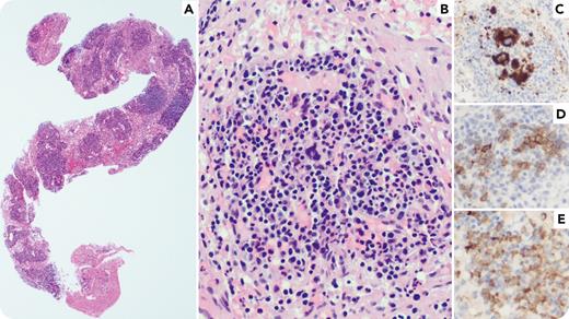
A 61-year-old man with a history of recurrent deep venous thromboses presented with dysphagia, a 30-lb (∼14-kg) weight loss in the past year, and fevers of unknown origin. A complete blood cell count showed macrocytic anemia (hemoglobin, 7.4 g/dL; mean corpuscular volume, 103 fL) with normal white blood cell (absolute neutrophil count, 2350/μL; absolute lymphocyte count, 550/μL, and absolute monocyte count, 90/μL) and platelet count. A positron emission tomography (PET) scan showed multiple areas of fluorodeoxyglucose (FDG) avidity, including the abdominal lymph nodes, facial/submandibular tissue, bone marrow, and spleen. A bone marrow biopsy showed vacuolation of myeloid and erythroid precursors with hypercellularity. A retroperitoneal lymph node biopsy showed lymphoid tissue separated by islands of nucleated red blood cells, myeloid precursors, and megakaryocytes (panels A-E; original magnifications ×50 [panel A] and ×200 [panel B], hematoxylin and eosin stain; ×200 CD61 [panel C]; ×200 E-cadherin [panel D]; and ×200 myeloperoxidase [panel E]), with no definitive evidence of a lymphoproliferative disorder. Subsequent genetic testing showed a UBA1 mutation (c.121A>G, p.Met41Val), consistent with VEXAS syndrome.
VEXAS syndrome is an X-linked somatic disorder caused by mutations of UBA1. Aside from the prototypic vacuoles in myeloid and erythroid precursors, it can lead to inflammatory and hematologic manifestations, including cytopenias. Extramedullary hematopoiesis (EMH) can be a cause of concerning FDG avidity on PET and has not previously been documented in this entity. Other causes of EMH, such as thalassemia, fibrosis, or a myeloproliferative neoplasm, were not present in this patient.
For additional images, visit the ASH Image Bank, a reference and teaching tool that is continually updated with new atlas and case study images. For more information, visit https://imagebank.hematology.org.


This feature is available to Subscribers Only
Sign In or Create an Account Close Modal