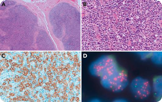
A 51-year-old Black woman presented with cervical lymphadenopathy. Lymph node biopsy showed effaced lymph node architecture with a nodular, mixed cellular infiltrate and bands of sclerosis (panel A; objective magnification 4×/total magnification ×40; hematoxylin and eosin [H&E] stain). Large malignant cells with multilobated nuclei were scattered in a background of small lymphocytes, histiocytes, plasma cells, and eosinophils (panel B; objective magnification 40×/total magnification ×400; H&E stain). The tumor cells expressed CD30 (panel C; objective magnification 40×/total magnification ×400), CD15, CD4, CD43, granzyme B, and TIA1. ALK, CD3, CD4, CD8, CD20, CD45, PAX5, and Epstein-Barr virus were negative. Polymerase chain reaction showed a clonal T-cell receptor gene rearrangement. Fluorescence in situ hybridization using a JAK2 break-apart probe showed separation of the 5′ (green) and 3′ (red) probes with 3′ amplification (panel D; objective magnification 100×/total magnification ×1000). DUSP22 rearrangement was absent. Sequencing showed an ATXN2L::JAK2 fusion transcript. ERBB4 was not expressed. The patient was treated with CHOP (cyclophosphamide, doxorubicin hydrochloride [hydroxydaunorubicin], vincristine sulfate [Oncovin], prednisone) chemotherapy followed by autologous stem cell transplantation and is alive and relapse free 105 months after diagnosis.
Awareness of anaplastic large cell lymphoma (ALCL) mimicking classic Hodgkin lymphoma (CHL) is essential to avoid misdiagnosis, particularly in ALK-negative (ALK–) cases. Accurate diagnosis is critical, as ALCL and CHL are typically treated with different chemotherapy regimens. JAK2 rearrangements are seen in ∼6% of CD30+/ALK– T-cell lymphomas. Fluorescence in situ hybridization or sequencing should be considered because JAK2 represents a potential therapeutic target in these cases.
For additional images, visit the ASH Image Bank, a reference and teaching tool that is continually updated with new atlas and case study images. For more information, visit http://imagebank.hematology.org.


This feature is available to Subscribers Only
Sign In or Create an Account Close Modal