

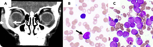
A four-year-old girl presented with an asymptomatic unilateral orbital swelling with no other abnormal physical findings. Her pediatrician obtained an MRI and referred her to ophthalmology to biopsy an orbital mass seen on MRI (panel A, arrow). Complete blood count (CBC) prior to biopsy showed anemia (Hgb 7.9 g/dL), thrombocytopenia (platelets 68 000 per µL), white blood cell count 5500 per μL, with 25% blasts (panel B, peripheral smear showing blast, arrow, and nearby lymphocyte, Wright-Giemsa stain (WG), original magnification ×1000). Peripheral blasts had lymphoblastic morphology and were positive by peripheral blood (PB) flow cytometry for CD10, CD19, CD22, TdT, and negative for light chains. Based on these findings, the diagnosis of B-acute lymphoblastic leukemia (B-ALL) was made, and the biopsy was canceled. Bone marrow aspiration showed sheets of blasts (panel C; 85.7%, WG, original magnification ×1000).
Clinical presentation with an extramedullary mass in pediatric B-ALL is an infrequent and often underrecognized association. Ten percent or less of orbital tumors in childhood are due to leukemias and lymphomas, with granulocytic sarcoma the most common underlying hematologic malignancy with an orbital mass presentation. This case illustrates this association and underscores the role of reviewing CBC and PB smear before performing a biopsy to optimize the initial investigations in these cases.
For additional images, visit the ASH Image Bank, a reference and teaching tool that is continually updated with new atlas and case study images. For more information, visit http://imagebank.hematology.org.

This feature is available to Subscribers Only
Sign In or Create an Account Close Modal