

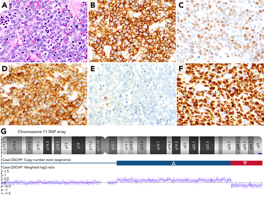
A 26-year-old-man presented to the hospital with abdominal pain. Abdominal imaging showed acute appendicitis, and the patient underwent an appendectomy. The appendectomy specimen showed extensive involvement of the wall of the appendix by a diffuse infiltrate of medium-sized atypical lymphocytes with abundant coarse apoptotic debris in the background, which imparted a “starry-sky” appearance to the infiltrate (panel A; hematoxylin and eosin stain, ×40 objective; total magnification ×400). By immunohistochemistry, the atypical lymphocytes expressed CD20 (panel B; ×40 objective; total magnification ×400), c-Myc (panel C; ×40 objective; total magnification ×400), and CD10 (panel D; ×40 objective; total magnification ×400) and lacked BCL2 (panel E; ×40 objective; total magnification ×400). In addition, the atypical cells expressed BCL6 and lacked TdT, CD34, MUM1, and EBER. Ki-67 showed a very high proliferation rate of >95% (panel F; ×40 objective; total magnification ×400). Fluorescence in situ hybridization (FISH) for a MYC rearrangement was negative, and single-nucleotide polymorphism (SNP) array revealed a proximal (centromeric) gain in segment 11q12.1-q23.3 and distal (telomeric) loss in segment 11q23.3-qter on the long arm of chromosome 11 (panel G; SNP-array results for chromosome 11). The case was diagnosed as Burkitt-like lymphoma with 11q aberration.
Burkitt-like lymphoma with 11q aberration is a relatively newly recognized entity that may mimic Burkitt lymphoma, including with expression of c-Myc. These cases diagnostically lack MYC rearrangements by FISH and show characteristic abnormalities of 11q.
For additional images, visit the ASH Image Bank, a reference and teaching tool that is continually updated with new atlas and case study images. For more information, visit http://imagebank.hematology.org.

This feature is available to Subscribers Only
Sign In or Create an Account Close Modal