

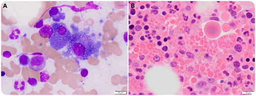
A 59-year-old man had an incidental finding of 0.8 g/dL immunoglobulin G (IgG) gamma monoclonal protein during an evaluation for Crohn’s disease. A bone marrow biopsy showed 5% plasma cells, and an observational approach was taken. Three years later, his M-protein level increased to 3.1 g/dL, and a repeat bone marrow biopsy showed progression to multiple myeloma with 50% plasma cells. The most striking feature was the increased number of Mott cells and extracellular Ig globules on the aspirate smears (panel A: Wright-Giemsa stain, ×60 oil objective, original magnification ×600) and in the trephine biopsy (panel B: hematoxylin and eosin stain, ×60 oil objective, original magnification ×600).
Mott cells have the classic “bunch of grapes” appearance of the plasma cell cytoplasm packed with Ig inclusions (Russell bodies). Mott cells can be seen in plasma cell dyscrasias and reactive plasmacytoses such as chronic inflammatory conditions, autoimmune-mediated diseases (eg, Hashimoto’s thyroiditis and rheumatoid arthritis), and rare conditions like Wiskott-Aldrich syndrome and von Recklinghausen’s neurofibromatosis. Ultrastructural studies have shown that the inclusions are made up of condensed Ig’s within vesicular structures derived from dilated endoplasmic reticulum cisternae. Other investigators have linked specific genetic loci to Mott cell formation and hypergammaglobulinemia.
For additional images, visit the ASH Image Bank, a reference and teaching tool that is continually updated with new atlas and case study images. For more information, visit http://imagebank.hematology.org.

This feature is available to Subscribers Only
Sign In or Create an Account Close Modal