

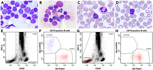
A 58-year-old woman with a history of low-grade non-Hodgkin lymphoma, chronic obstructive pulmonary disease, non-ST-elevation myocardial infarction, and adenocarcinoma of the lung presented with an increasing mass in a routine chest X-ray. Follow-up positron emission tomography and computed tomography showed lymphadenopathy. Lung and lymph node biopsies and a bone marrow aspirate were performed to investigate possible lymphoma. Microscopic investigation of the bone marrow smears showed increased lymphocytic proliferation and sheets of atypical lymphocytes with indented nuclei (panels A-B; ×100 objective, original magnification ×1250; May-Grünwald-Giemsa stain). In the peripheral blood smear, similar atypical lymphocytes were seen (panels C-D; ×100 objective, original magnification ×1250; May-Grünwald-Giemsa stain). Flow cytometry analysis of the bone marrow aspirate showed a small monoclonal B-cell population expressing CD10, CD19, CD20, CD22, CD79b, and surface immunoglobulin κ but lacking CD45 (panels E-H). The CD45 antigen is normally universally expressed in variable intensities on all leukocytes. Usually, this marker is used to gate out non-nucleated cells. In this case, however, the CD45-negative follicular lymphoma cells were almost overlooked using the normal gating strategy. Gating on all cells was necessary to find the monoclonal B-cell population and diagnose the patient with follicular lymphoma.
This rarely reported finding shows that although flow cytometry can be extremely useful for the diagnosis of hematological disorders, one should always be aware of rare, atypical immunophenotypes.
For additional images, visit the ASH Image Bank, a reference and teaching tool that is continually updated with new atlas and case study images. For more information, visit http://imagebank.hematology.org.

This feature is available to Subscribers Only
Sign In or Create an Account Close Modal