

Current understanding of acute myeloid leukemia (AML) assumes a developmental hierarchy, in which a minor fraction of primitive and quiescent leukemia stem cells (LSC) sustain clonal propagation of disease. These therapy-resistant LSC may be the basis of relapse, as supported by data correlating the presence of LSC gene expression signatures at diagnosis with poor prognosis (Ng et al, Nature 2016). Novel approaches tracing LSC fates at single cell level before, during and after chemotherapy (CTX) are needed to confirm their biological relevance and derive new, LSC-focused diagnostic and therapeutic strategies.
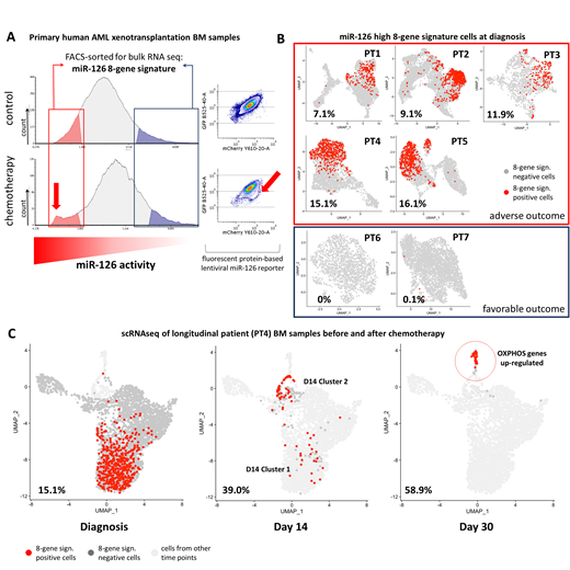
We and others have previously linked key LSC properties, such as quiescence and therapy resistance, to complex transcriptional regulation orchestrated by miR-126 (Lechman et al, Cancer Cell 2016). Furthermore, we provided proof of concept that LSCs could be prospectively isolated as miR-126(high) cells exploiting a lentiviral reporter vector capturing miR-126 bioactivity in live cells with single cell resolution. To extend these studies, we transduced primary blasts from n=3 AML patients (pts) carrying NPM1 and FLT3-ITD mutations with the miR-126 reporter, followed by xenografting (PDX). Blasts showed intra-tumor heterogeneity (ITH) in terms of miR-126 activity, with a minor fraction identified as miR-126(high). Limiting dilution secondary transplantation of FACS sorted miR-126(high) and -(low) blasts proved strong enrichment of repopulating activity within the miR-126(high) compartment in all 3 pts. On the contrary, no LSC enrichment could be verified in the CD34+CD38- fraction in 1 patient, suggesting that high miR-126 activity represents a more robust LSC identifier than commonly used surface markers. Next, we investigated the impact of daunorubicin and cytarabine CTX on miR-126-reporter+ blasts (n=3 AML) in PDX. Surprisingly, overall miR-126 activity diminished in post CTX residual AML compared to controls, compatible with a loss of blast quiescence. CTX accentuated ITH by uncovering a subset of blasts with very high levels of miR-126, distinct from the bulk population, which may correspond to residual quiescent LSC (Fig A). We then performed bulk RNA sequencing on miR-126(high) and miR-126(low) subsets from CTX and control PDX. While miR-126(low) blasts from both groups expressed markers of myeloid differentiation, miR-126(high) blasts were enriched for published hematopoietic stem cell hallmark signatures. Integrating differentially expressed genes between miR-126(high) and-(low) subsets at steady state and post CTX, we extrapolated a novel 8 gene signature associated with miR-126(high) blasts. Of note, patients from the AML TCGA PanCancer Atlas Cohort (n=161) harboring overexpression in one or more of these 8 genes had significantly decreased overall survival (10 vs 19 months, Logrank test p-value = 0.018).
To further test whether our 8-gene miR-126(high) signature reveals ITH in patients, we performed single cell RNA sequencing (scRNAseq) of AML patient BM aspirates at diagnosis (n= 6). Blasts were identified based on the detection of mutated NPM1 transcripts in single cells. In 5 out of 7 patients expression of the miR-126 signature mapped to specific clusters of blasts identified by unsupervised shared nearest neighbor algorithm, confirming that it identifies ITH in patient samples. Interestingly, we detected miR-126 signature(high) blasts in pts with poor prognosis (n=4) and not in those with favorable outcome (n=2) (Fig B).
To investigate ITH across longitudinal samples, we next performed scRNAseq of residual AML from a representative patient assessed early after CTX. In line with our PDX CTX model displaying increased miR-126 ITH, blasts on day14 of induction CTX segregated into 2 different clusters: cluster 1 containing LSC-like cells with miR-126(high) signature and similar transcriptional profile to the diagnosis counterpart; cluster 2, instead, was composed of actively cycling blasts. Residual blasts at day30, in addition to uniformly expressing the miR-126(high) signature, differed from diagnosis and day14 blasts by displaying cell cycle quiescence and induction of oxidative phosphorylation genes (Fig C). In summary, we have set up and applied PDX LSC modeling to clinically relevant patient samples to address AML intra-tumor heterogeneity and to pinpoint novel relevant transcriptomic features of LSC at diagnosis and after chemotherapy.
Gentner:Genenta Science: Consultancy, Equity Ownership, Research Funding.

This feature is available to Subscribers Only
Sign In or Create an Account Close Modal