

The defective necroptotic pathway and cytokines were important in the pathogenesis and progression of chronic lymphocytic leukemia (CLL). On the other hand, selenite could induce necroptosis in various sold malignancies by generating reactive oxygen species. However, the relationship between necroptosis and cytokines still remains unclear. Here, we managed to clarify the role of different cytokines and selenite in the defective necroptotic pathway of CLL.
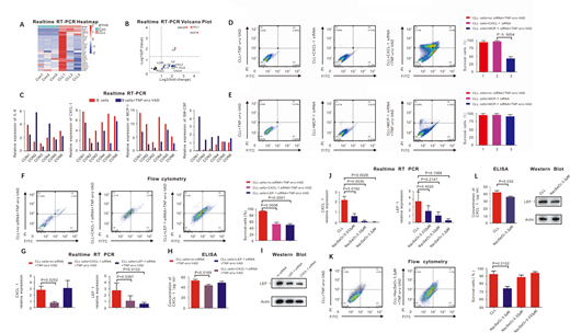
We first screened the expression of different cytokines related to malignancies between CLL patients and controls using Realtime RT-PCR. Only the expression of CXCL-1, MCP-1, IL-6 and GM-CSF was significantly higher in CLL patients than that of controls. (Figure A, B) In normal B lymphocytes, TNF-α and z-VAD could induce necroptosis and also downregulated the expression of CXCL-1 and MCP-1, which indicated that CXCL-1 and MCP-1 might have correlation with necroptosis. (Figure C) After knockdown of CXCL-1 rather than MCP-1 by siRNA, CLL cells restored the TNF-α/z-VAD induced necroptosis measured by flow cytometry. (Figure D, E)
To assess the association between CXCL-1 and lymphoid enhancer-binding factor 1 (LEF-1), which has already been confirmed as the key protein in the necroptotic pathway of CLL, we first performed flow cytometry to verify that CLL cells restored TNF-α/z-VAD induced necroptosis after inhibition of either CXCL-1 or LEF-1. (Figure F) Then, we used Realtime RT-PCR, Western Blot and ELISA to confirm that the expression of LEF-1 was downregulated after inhibiting the expression of CXCL-1 by siRNA, however, the expression of CXCL-1 did not change significantly after knockdown of LEF-1. (Figure G, H, I) This results demonstrated that CXCL-1 located the upstream of LEF-1.
To figure out the relationship between cytokines, necroptosis and selenite, we first construct the selenite concentration gradient and selenite with different concentrations was added into CLL cells together with TNF-α and z-VAD. We found that selenite could downregulate the expression of CXCL-1 but had little influence on LEF-1 measured by Realtime RT-PCR. (Figure J) Then, flow cytometry was performed to calculate the percentage of survival CLL cells and only 3.2μM selenite could significantly induce necroptosis of CLL cells. (p = 0.0102, Figure K) Finally, both Western Blot and ELISA showed the similar result that 3.2μM sodium selenite downregulated the translational expression of CXCL-1 but had little impact on LEF-1. (Figure L)
No relevant conflicts of interest to declare.

This feature is available to Subscribers Only
Sign In or Create an Account Close Modal