

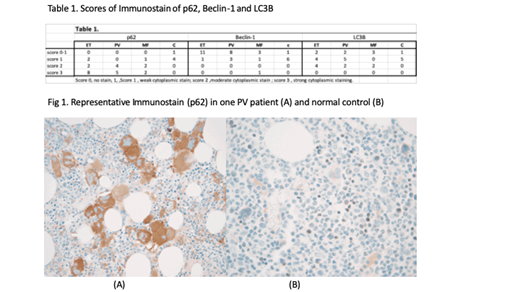
We previously reported that autophagy was defective in classical Philadelphia negative (Ph -) Myeloproliferative Neoplasm (MPN) by demonstrating increased p62, decreased Beclin-1 by RT-PCR and Western Blot (WB) methods and decreased LC3-II by WB (ASH annual meeting poster 2018) in peripheral blood mononuclear cells. Now, we further performed immunohistochemical staining of these autophagy markers on the bone marrow biopsy specimens. Methods: Formalin-fixed and paraffin-embedded bone marrow samples were stained with primary antibodies against Beclin-1 (mouse monoclonal, Millipore), LC3B (rabbit monoclonal, Cell Signaling Technology), and p62 (mouse monoclonal, Cell Signaling Technology). The tissue sections were analyzed using VENTANA BenchMark Ultra System (Ventana Medical Systems, Inc.) according to the manufacturer's protocol. Patients who were studied included 12 essential thrombocythemia (ET), 10 polycythemia (PV), and 5 myelofibrosis (MF) (including 1 post-ET MF and 1 post-PV MF). Scoring was given based on the degree of cytoplasmic staining. Score 1 included weak cytoplasmic stain, score 2 included moderate cytoplasmic stain and score 3 was given for strong cytoplasmic staining . Results: 1) As shown in Fig1, the predominant cells which stained positive including p62, Beclin-1 , or LC3 B were mostly found on the megakaryoctes, although very few of other cell types were found to have positive staining as well 2) As shown in Table 1, the immunostaining results were in general correlated to the RT-PCR , or western blot findings that a defective autophagy was demonstrated in MPN patients with combined finding of a stronger staining in p62,and weak staining in Beclin-1 and LC3B. Conclusion: We have demonstrated defective autophagy in these Ph (-) MPN, by immunostaining of the bone marrow specimens, and demonstrating positivity for defective autophagy predominantly in megakaryocytes. Since megakaryocytes are the most important cells involved in the pathogenesis of MPN, we propose that defective autophagy in megakaryocytes play an important role in the pathogenesis of these diseases.
Wang:Incyt: Research Funding.

This feature is available to Subscribers Only
Sign In or Create an Account Close Modal