

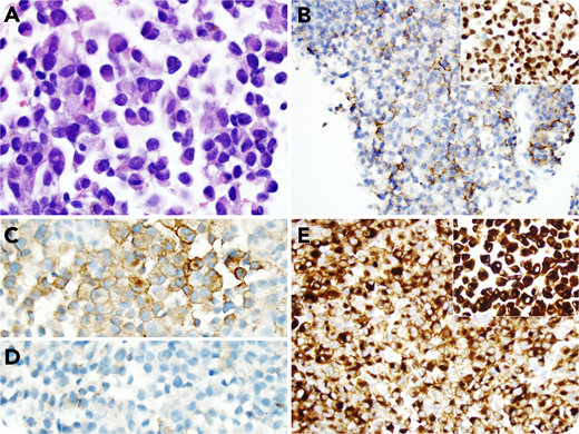
A 63-year-old woman with a history of immunoglobulin A κ–restricted plasma cell myeloma, which relapsed after autologous hematopoietic stem cell transplantation, was recently noted to have adenopathy in the right periauricular area. A core needle biopsy was performed, which showed sheets of cells with eccentric nuclei, eosinophilic granular cytoplasm, round nuclear contours, and clumped chromatin with prominent red nucleoli, characteristic of plasma cells (panel A; hematoxylin and eosin stain; objective 100×; original magnification ×1000). Some cells showed nuclear inclusions reminiscent of Dutcher bodies. Immunostains showed these cells were variably positive for CD138 and positive for MUM1 (panel B and inset, respectively; objective 20×; original magnification ×200) and unusually had only variable surface κ light chain expression (panel C; objective 40×; original magnification ×400), whereas λ was negative (panel D; objective 40×; original magnification ×400). Because of the unusual staining pattern, further investigation was performed, which revealed the patient had a history of melanoma with an unknown primary site. Immunostaining with HMB-45 and Melan-A showed diffuse staining (panel E and inset, respectively; objective 20×; original magnification ×200), consistent with metastatic melanoma.
Melanoma is known to mimic the histology of many tumors, and this case underlines that it can also mimic plasma cells. MUM1 is frequently expressed in melanoma, but the unusual staining pattern seen in this case should prompt the pathologist to further investigate the clinical history and expand the immunostain workup.
For additional images, visit the ASH Image Bank, a reference and teaching tool that is continually updated with new atlas and case study images. For more information, visit http://imagebank.hematology.org.

This feature is available to Subscribers Only
Sign In or Create an Account Close Modal