

A 22-year-old woman was admitted to our hospital for management of respiratory failure due to parainfluenza virus infection and aspiration, requiring noninvasive ventilation. She had a history of Sanfilippo disease (mucopolysaccharidosis type 3B; diagnosed at a different institution) that manifested clinically with severe intellectual disability, chronic respiratory disease with suppurative bronchitis, seizure disorder, obstructive sleep apnea, scoliosis, and required gastrostomy feeding.
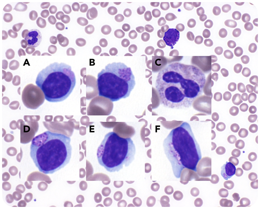
A peripheral blood film was performed during this admission that showed a leukoerythroblastic picture, with nucleated red cells and left-shifted myelopoiesis, all features of bone marrow stress. The lymphocytes displayed cytoplasmic vacuoles, as well as azurophilic granules surrounded by halos, called Alder-Reilly anomaly, in keeping with Sanfilippo syndrome (panels A-F; Wright-Giemsa stain, original magnification ×1000, background image original magnification ×400). The differentials of the lymphocyte features include ehrlichiosis and large granular lymphocytes, with the latter displaying azurophilic granules that are often slightly smaller than those found in the mucopolysaccharidosis syndromes and are not surrounded by halos.
A 22-year-old woman was admitted to our hospital for management of respiratory failure due to parainfluenza virus infection and aspiration, requiring noninvasive ventilation. She had a history of Sanfilippo disease (mucopolysaccharidosis type 3B; diagnosed at a different institution) that manifested clinically with severe intellectual disability, chronic respiratory disease with suppurative bronchitis, seizure disorder, obstructive sleep apnea, scoliosis, and required gastrostomy feeding.
A peripheral blood film was performed during this admission that showed a leukoerythroblastic picture, with nucleated red cells and left-shifted myelopoiesis, all features of bone marrow stress. The lymphocytes displayed cytoplasmic vacuoles, as well as azurophilic granules surrounded by halos, called Alder-Reilly anomaly, in keeping with Sanfilippo syndrome (panels A-F; Wright-Giemsa stain, original magnification ×1000, background image original magnification ×400). The differentials of the lymphocyte features include ehrlichiosis and large granular lymphocytes, with the latter displaying azurophilic granules that are often slightly smaller than those found in the mucopolysaccharidosis syndromes and are not surrounded by halos.
For additional images, visit the ASH Image Bank, a reference and teaching tool that is continually updated with new atlas and case study images. For more information, visit http://imagebank.hematology.org.

This feature is available to Subscribers Only
Sign In or Create an Account Close Modal