

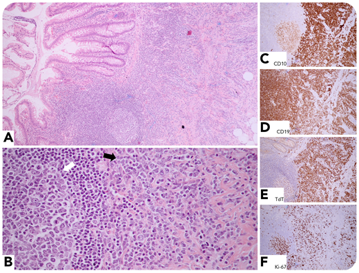
A 29-year-old man with a history of Philadelphia chromosome–positive B-lymphoblastic leukemia/lymphoma (B-ALL/LBL), 3 months post–allogeneic stem cell transplantation, presented with right lower quadrant abdominal pain, diarrhea, nausea, and vomiting. Computed tomography scan was suspicious for appendicitis, and an appendectomy was performed. The histologic sections showed circumferential leukemic infiltrate (panel A; original magnification ×40, hematoxylin and eosin stain). Remnants of gut-associated lymphoid tissue (white arrow) were noted adjacent to the neoplasm (black arrow) (panel B; original magnification ×200, hematoxylin and eosin stain). The neoplastic cells expressed CD10 (panel C; original magnification ×40, immunohistochemical stain), CD19 (panel D; original magnification ×40, immunohistochemical stain), and focal CD34, CD79a, PAX5, and TdT (panel E; original magnification ×40, immunohistochemical stain); they were nonreactive with CD2, CD3, CD20, and MPO. The germinal centers and the neoplasm were positive for CD10, but only the latter was TdT positive. Ki-67 labeling index was 50% (panel F; original magnification ×40, immunohistochemical stain).
B-ALL/LBL is an uncommon disease, with a global annual incidence rate of 1 to 4.75 cases per 100 000. Three fourths of the cases occur in children younger than 6 years old. Appendiceal involvement has been reported but is extremely rare.
For additional images, visit the ASH Image Bank, a reference and teaching tool that is continually updated with new atlas and case study images. For more information, visit http://imagebank.hematology.org.

This feature is available to Subscribers Only
Sign In or Create an Account Close Modal