

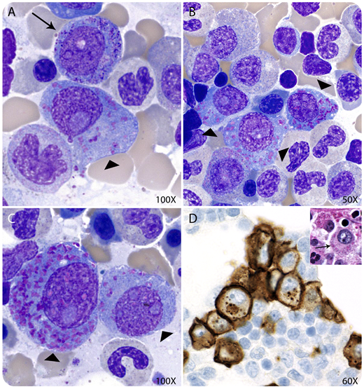
A 70-year-old man with a history of treated immunoglobulin G (IgG)–κ plasma cell myeloma was referred to our institution for continuation of care. A preliminary differential cell count demonstrated 10% promyelocytes and 2% plasma cells. The second review of Wright-stained bone marrow aspirate smears showed 12% atypical plasma cells and 1% promyelocytes (panel A, arrow). Curiously, most plasma cells contained a variable number of coarse cytoplasmic azurophilic crystalline inclusions and resembled promyelocytes (panels A-C, arrowheads). A core biopsy demonstrated ∼15% plasma cells with granular cytoplasm (panel D inset, arrow), negative for periodic acid-Schiff, positive for CD138 by immunohistochemistry (panel D), and κ light chain restricted by in situ hybridization.
Plasma cell cytoplasmic azurophilic crystalline inclusions are rare. They include azurophilic granules of variable size and shapes and Auer rod–like crystals. In the current case, the morphologic features in the plasma cells were prominent enough to confuse them with promyelocytes (panels A-C), but ancillary tests confirmed their plasma cell origin. Azurophilic crystalline inclusions are thought to represent abnormal immunoglobulin deposits secondary to a block in the protein synthetic pathway, whereas Auer rod–like crystals appear to have a lysosomal origin. The recognition of plasma cell cytoplasmic azurophilic inclusions is crucial to avoid erroneous differential cell counts or a misdiagnosis of acute myeloid leukemia.
For additional images, visit the ASH Image Bank, a reference and teaching tool that is continually updated with new atlas and case study images. For more information, visit http://imagebank.hematology.org.

This feature is available to Subscribers Only
Sign In or Create an Account Close Modal