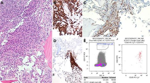
A 57-year-old man underwent bone marrow examination to investigate lumbar compression fractures and an elevated λ light chain level of 3508 mg/L. The marrow aspirate demonstrated 50% abnormal plasma cells diagnostic of myeloma. The trephine biopsy revealed an extensive infiltrate of plasma cells, as well as a paratrabecular aggregate of spindle-shaped cells (panel A; hematoxylin and eosin stain, original magnification ×400). These latter cells were not positive for CD138 (panel B; original magnification ×400) but stained positive for both mast cell tryptase (panel C; original magnification ×400) and CD117 (panel D; original magnification ×400). Immunophenotyping confirmed these as aberrant (using CD117 vs side scatter gating strategy) with expression of CD2 and CD25 (panel E). The combination of the histologic appearance (major criterion) and antigenic aberrancy (minor criterion) fulfilled the diagnostic criteria of systemic mastocytosis. Apart from back pain attributable to the compression fractures, there were no other symptoms of mastocytosis present. A CKIT D816V mutation (variant allele fraction <0.1%) was detected; however, serum tryptase level was not elevated.
“Systemic mastocytosis with an associated hematologic neoplasm” (2016 World Health Organization update) is most frequently found with myeloid malignancies. Myeloma has rarely been described as an association in this category. Initially, the spindle-shaped cell aggregate of mast cells was thought to be part of the plasma cell infiltrate, demonstrating the importance of confirmatory immunohistochemistry and immunophenotyping in resolving cases with atypical morphology.
For additional images, visit the ASH Image Bank, a reference and teaching tool that is continually updated with new atlas and case study images. For more information, visit http://imagebank.hematology.org.


This feature is available to Subscribers Only
Sign In or Create an Account Close Modal