To the editor:
Double cord blood (CB) transplantation (dCBT) is an accepted treatment of patients with hematologic malignancies.1,2 In the vast majority of dCBT recipients, 1 unit emerges as the sole source of long-term hematopoiesis.3 As measured by standard clinical testing for chimerism (usually by short-tandem-repeat [STR] polymorphism), the “losing” unit usually becomes undetectable within the first month after transplantation.4-7 However, anecdotal cases in which the losing unit reemerges and contributes to hematopoiesis suggest long-term persistence of the losing unit in a quiescent state.
Although studies suggest an in vivo immune-mediated mechanism for single-donor dominance, there is no established evidence that the losing unit is definitively rejected.6-8 Moreover, limited data suggest that mixed-unit chimerism (the persistence of both donor CB units) may be associated with a potentially advantageous, enhanced graft-versus-tumor effect.9 Indeed, coexistence of semiallogeneic cells in the same individual is already a well-recognized natural phenomenon (microchimerism) resulting from bidirectional maternal-fetal exchange during pregnancy with persistence in respective individuals decades later.10 Naturally acquired microchimerism is found in healthy individuals, in organs and circulation, without apparent graft-versus-host reaction or graft rejection and has been associated with both health benefits and risks, pointing to functional capacity.10,11
We hypothesized that a similar phenomenon also occurs after dCBT more commonly than would be suggested by estimates of mixed-unit chimerism by standard clinical measures, with “occult” presence of cells derived from the losing unit in the clinical setting of complete single CB unit dominance after dCBT. Using a sensitive technique developed for microchimerism analysis, we sought to determine whether very low levels of the losing unit could be identified.
Bone marrow (BM) and peripheral blood (PB) samples were collected at approximately days 28, 80, and 365 after transplant for clinical chimerism testing using a standard STR approach, and residual blood specimens were stored for research. PB mononuclear cells (PBMCs) were collected by density-based centrifugation. Cell-lineage subsets (CD3+, CD33+, and CD56+) were isolated by fluorescence-activated cell sorting at the time of the blood draw.
HLA-genotyping data for subjects and CB units were reviewed to identify an HLA polymorphism unique to the losing CB unit to target using a panel of HLA-specific quantitative polymerase chain reaction (QPCR) assays. All assays were developed to detect the DNA equivalent of 1 cell in 20 000. The approach provides a standardized method for microchimerism testing that is highly sensitive and highly specific as previously described.12,13 DNA extracted from available BM, PBMC, and PB cell subset samples was tested with an HLA-specific QPCR assay unique to the losing unit. The total genome equivalent (GEq) tested median amounts were 9.5 × 10⁴ (range, 1.1 × 10⁴ to 47.0 × 10⁴) for PBMCs, 11.6 × 10⁴ (range, 1.5 × 10⁴ to 16.7 × 10⁴) for BM, and 2.1 × 10³ (range, 2.5 × 10² to 3.3 × 10⁴) for cell subsets.
Among consecutive patients undergoing a myeloablative CBT on protocols NCT00719888 and NCT00796068 between 2006 and 2014, we selected 14 patients who received a dCBT with either high-dose total-body irradiation (TBI)-based conditioning consisting of 1320 cGy TBI, fludarabine 75 mg/m2, and cyclophosphamide 120 mg/kg (n = 8), or low-dose TBI-based conditioning with 200 cGy TBI, treosulfan 42 mg/m2, and fludarabine 150 mg/m2 (n = 6). All patients received cyclosporine plus mycophenolate mofetil for acute graft-versus-host disease (GVHD) prevention. Results were analyzed according to detection or not of the losing unit, and quantitative results summarized and expressed as DNA GEq number of cells from the losing unit per total GEq of DNA tested. All study activities were approved by the Fred Hutchinson Cancer Research Center Institutional Review Board, and all participants provided written informed consent in accordance with the principles of the Declaration of Helsinki.
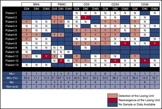
Subjects were selected according to demonstration of single CB unit dominance by clinical testing (n = 13, subjects), with 1 stable mixed unit-unit chimerism included (n = 1). The median age at transplant was 32 years (range, 10-62 years) and the median weight was 72.2 kg (range, 32.5-107 kg). Pretransplant diseases included acute myeloid leukemia (n = 10), acute lymphoblastic leukemia (n = 3), and myeloproliferative disorder (n = 1). No patients experienced primary graft failure. For the overall study population (n = 14), the median time to neutrophil engraftment was 23 days (range, 14-44 days). Of the 13 subjects with single-unit dominance, absence of the losing unit by clinical STR testing was confirmed at a median time of 20 days (range, 11-28 days). The patient with clinical mixed-unit chimerism gave positive results as expected. QPCR testing results are summarized in Figure 1. Clinical characteristics of the 13 patients are shown in Table 1.
Detection and reemergence of the losing unit in 13 patients with single-donor dominance in whole BM in PBMCs and in CD3, CD33, and CD56 cell subsets at days 28, 80, and 365 posttransplant. BMA, bone marrow aspiration; Mc−, negative microchimerism; Mc+, positive microchimerism; Non-eval, not evaluable.
Detection and reemergence of the losing unit in 13 patients with single-donor dominance in whole BM in PBMCs and in CD3, CD33, and CD56 cell subsets at days 28, 80, and 365 posttransplant. BMA, bone marrow aspiration; Mc−, negative microchimerism; Mc+, positive microchimerism; Non-eval, not evaluable.
Patient characteristics
| Patient no. . | Sex . | Age, y . | Disease . | HLA units to patient . | HLA unit to unit . | TNC, ×107/kg . | CD34, ×105/kg . | ANC, d . | aGVHD grade . | Relapse day* . | Death day* . |
|---|---|---|---|---|---|---|---|---|---|---|---|
| 1 | F | 14 | AML | 5/6 and 5/6 | 4/6 | 7.9 | 4.5 | 14 | 3 | 1378 | 1471 |
| 2 | F | 21 | ALL | 4/6 and 4/6 | 3/6 | 8.1 | 3.0 | 26 | 2 | 138 | |
| 3 | F | 50 | AML | 5/6 and 4/6 | 4/6 | 5.3 | 2.4 | 19 | 2 | ||
| 4 | F | 24 | CML | 4/6 and 4/6 | 4/6 | 5.3 | 2.2 | 44 | 2 | ||
| 5 | M | 39 | AML | 4/6 and 4/6 | 3/6 | 4.8 | 2.1 | 25 | 2 | ||
| 6 | M | 62 | AML | 4/6 and 5/6 | 3/6 | 5.5 | 2.8 | 17 | 0 | 203 | 229 |
| 7 | F | 14 | AML | 5/6 and 5/6 | 4/6 | 3.8 | — | 19 | 2 | ||
| 8 | M | 10 | AML | 4/6 and 4/6 | 4/6 | 14.3 | — | 25 | 3 | ||
| 9 | M | 52 | ALL | 4/6 and 4/6 | 4/6 | 5.8 | 4.2 | 18 | 2 | ||
| 10 | M | 25 | AML | 4/6 and 4/6 | 4/6 | 3.8 | 1.4 | 36 | 2 | 410 | |
| 11 | F | 51 | AML | 5/6 and 5/6 | 4/6 | 4.2 | 1.7 | 20 | 2 | ||
| 12 | M | 13 | ALL | 5/6 and 4/6 | 3/6 | 4.6 | 2.9 | 32 | 1 | ||
| 13 | M | 42 | AML | 4/6 and 4/6 | 4/6 | 5.1 | 4.4 | 32 | 0 |
The day of neutrophil engraftment was defined as the first of 3 consecutive days of an ANC of ≥0.5 × 10⁹/L or greater.
aGVHD, acute GVHD; ALL, acute lymphoblastic leukemia; AML, acute myeloid leukemia; ANC, absolute neutrophil count; CML, chronic myelogenous leukemia; TNC, total nucleated dose.
Blank entries indicate that the event of interest did not happen in those patients.
Results showed detection of the losing unit in 8 of 13 subjects (62%) for at least 1 time point and at least 1 sample type. In PBMCs, 4 of 7 (57%), 4 of 7 (57%), and 0 of 4 (0%) evaluable patients showed losing-unit detection at days 28, 80, and 365, respectively. In the CD3 subset, 4 of 10 (40%), 5 of 10 (50%), and 3 of 9 (33%) showed losing-unit detection at days 28, 80, and 365, respectively. In the CD33 subset, 6 of 11 (55%), 4 of 11 (36%), and 1 of 7 (14%) showed losing-unit detection at days 28, 80, and 365, respectively. In the CD56 subset, 4 of 11 (36%), 1 of 7 (14%), and 1 of 6 (17%) showed losing-unit detection at days 28, 80, and 365, respectively. In the BM samples, 5 of 10 (50%), 1 of 9 (11%), and 1 of 8 (13%) showed losing-unit detection at days 28, 80, and 365, respectively. Of samples that had losing-unit detection, the median losing-unit concentration, expressed and standardized per 105 total GEq, was 4.0 (range, 0.1 to 2.8 × 10⁴) for PBMCs, 4.2 (range, 0.1 to 1.5 × 10³) for BM, and 945.9 (range, 5.4 to 7.5 × 10³) for cell subsets. Overall results of the current study indicate that the losing CB unit, undetectable by STR testing, was still identifiable in the majority of patients following dCBT (Figure 1).
To date, no factors reliably predict single-unit dominance or the coexistence of both units.5,7,8 Several reports have shown persistence for varying time periods of both CB units as stable, mixed-unit chimerism, with potential influence on clinical outcomes suggested by limited data.9 Dominance reversion is another uncommon situation in which cells of the predominating CB unit gradually decline giving up dominance to the other unit in the state of mixed-unit chimerism.9 The mechanism of dominance is multifactorial involving intrinsic features of the CB units, graft-versus-graft immune interactions mediated by T cells, as well as graft-versus-host immune interactions.4,6,8 In our study, the losing unit was still present even after the dominance phenomenon was well established clinically in the majority of dCBT patients. The presence of cells from the losing unit in the CD3 component suggests the possibility of dynamic regulation despite an apparent quiescent stable state.
Limited data suggest mixed-unit chimerism may confer a heightened graft-versus-leukemia benefit underscoring the need for better and more sensitive approaches to chimerism testing to further understanding of the biology and clinical implications of losing-unit persistence.9 Similarly, the use of this sensitive technique could be of clinical interest in other settings such as the HLA-mismatched stem cell microtransplantation14 and after haplo-cord transplantation15 where the persistence of donor cells is thought to be even more transitory.
Additional larger and prospective studies incorporating this type of approach are needed to assess losing-unit detection for correlation with clinical outcomes. Understanding the immunological interfaces for the patient, the dominant, and the “losing” unit may also yield insights into mechanisms of GVHD and graft-versus-leukemia responses.
Authorship
Acknowledgments: The authors are grateful to the patients and families who consented to the use of clinical research results.
This work was supported by National Institutes of Health National Heart, Lung, and Blood Institute grant R01 HL117737.
Contribution: F.M., J.L.N., and C.D. participated in the study design and interpretation of data for the manuscript; H.G., D.C.O., and S.B.K. performed and analyzed the PCR data which were validated by J.L.N.; F.M. wrote the first draft; and H.G., D.C.O., S.B.K., J.L.N., and C.D. provided revisions and critical review of the final manuscript.
Conflict-of-interest disclosure: The authors declare no competing financial interests.
Correspondence: Filippo Milano, Clinical Research Division, Fred Hutchinson Cancer Research Center, 1100 Fairview Ave N, Mail Box MD-B306, Seattle, WA 98109; e-mail: fmilano@fredhutch.org.


This feature is available to Subscribers Only
Sign In or Create an Account Close Modal