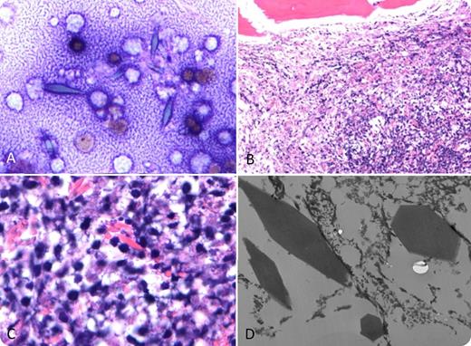
A 49-year-old man presented with a 3-month history of generalized body pain, 40-lb weight loss, night sweats, fevers, chills, and osteolytic lesions. Complete blood count showed anemia (hemoglobin of 7.5 g/dL); there was no eosinophilia. Bone marrow aspirate revealed blue-gray crystals (panel A; May-Grünwald-Giemsa stain, original magnification ×50). The biopsy revealed a cellular bone marrow with eosinophilic crystals and focal cell necrosis. Large crystals were visualized at higher magnification (panels B-C; hematoxylin and eosin stain, original magnifications ×10 and ×100, respectively). Immunohistochemical studies were negative for CD138, CD20, CD3, CD15, CD68, CD45, κ/λ chains, CD1A, CD79a, and CD38. Electron microscopy confirmed the bipyramidal structure longitudinally and in cross-section (panel D; original magnification ×74 500). Positron emission tomography/computed tomography revealed lymphadenopathy, and biopsy led to a diagnosis of T-cell lymphoblastic lymphoma.
These crystals are compatible with Charcot-Leyden crystals (CLCs), which are classically seen not only in the setting of eosinophilic inflammatory reactions but also in hematological malignancies in association with eosinophilia. CLCs have been described with bone marrow necrosis without eosinophilia in acute myelogenous leukemia but not previously in T-cell lymphoblastic lymphoma. This case emphasizes that the presence of CLCs in the setting of bone marrow necrosis can be associated with multiple hematologic malignancies.
For additional images, visit the ASH IMAGE BANK, a reference and teaching tool that is continually updated with new atlas and case study images. For more information visit http://imagebank.hematology.org.


This feature is available to Subscribers Only
Sign In or Create an Account Close Modal