In this issue of Blood, Beck et al1 describe adenosine diphosphate (ADP)–induced responses in the platelet phosphoproteome, which are overridden by the prostacyclin analog iloprost, revealing a hidden complexity of signal transduction that determines functional outcome.
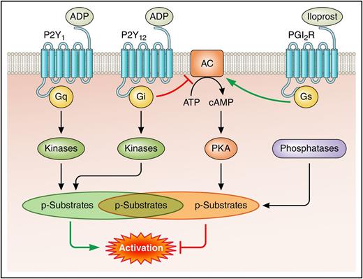
The study by Beck et al reveals a dynamic pool of phosphorylation events downstream of the ADP receptors P2Y1 and P2Y12 that are counteracted by the prostaglandin I2 (PGI2) analog iloprost, which signals via protein kinase A (PKA). Phosphatases are shown to play a central role in regulating these phosphorylation events; however, biochemical links to the receptors remain ambiguous. AC, adenylate cyclase; ATP, adenosine triphosphate; cAMP, cyclic adenosine monophosphate; PGI2R, prostaglandin I2 receptor. Professional illustration by Patrick Lane, ScEYEnce Studios.
The study by Beck et al reveals a dynamic pool of phosphorylation events downstream of the ADP receptors P2Y1 and P2Y12 that are counteracted by the prostaglandin I2 (PGI2) analog iloprost, which signals via protein kinase A (PKA). Phosphatases are shown to play a central role in regulating these phosphorylation events; however, biochemical links to the receptors remain ambiguous. AC, adenylate cyclase; ATP, adenosine triphosphate; cAMP, cyclic adenosine monophosphate; PGI2R, prostaglandin I2 receptor. Professional illustration by Patrick Lane, ScEYEnce Studios.
Platelet activation by ADP involves 2 receptors, P2Y1 and P2Y12, the importance of the latter exemplified by the clinical success of P2Y12 inhibitors in protecting against thrombosis.2 P2Y1 is a Gq-coupled receptor that signals predominantly through phospholipase Cβ and protein kinase C, resulting in platelet shape change and transient aggregation (see figure). P2Y12 is a Gi-coupled receptor that signals by inhibiting membrane-bound AC, thus reducing intracellular cAMP levels, and also by activating phosphoinositide 3-kinase.3 Collectively, these signals synergize with other activation signals to enhance the platelet response. However, a “black box” in the field is the question “Which other kinases are activated downstream of P2Y1 and P2Y12 and what are their substrates?” By using an unbiased phosphoproteomics-based approach, Beck et al uncover a wealth of new information about potential kinases, phosphatases, and substrates that drive ADP-triggered platelet activation. Interestingly, they found that the vast majority of regulated phosphorylation events, measured as either an increase or a decrease of enriched phophopeptides, took place in the first 10 seconds of stimulation, pointing to very rapid regulation. It should be noted that this includes serine, threonine, and tyrosine phosphorylated residues. Almost half of these early phosphorylation events were sustained, whereas the rest rapidly returned to basal levels. As was expected, many of the modified proteins participate in cytoskeletal remodeling, calcium signaling, secretion, aggregation, and other known platelet responses; however, the results also emphasize a striking number of kinases, guanosine triphosphate hydrolase enzyme (GTPase) exchange factors and GTPase-activating proteins, involved in these processes. The rapid and prominent dephosphorylation of many proteins after an initial burst in phosphorylation highlighted the central role of phosphatases in ADP-dependent platelet activation. As phosphatases emerge as critical regulators of platelet activation, these novel findings warrant further investigation.4
Endothelium-derived prostacyclin, also referred to as PGI2, is one of the most powerful inhibitors of platelet activation. PGI2 signals via the Gs-coupled PGI2 receptor, which stimulates AC activity, causing a rise in intracellular cAMP levels and activation of PKA (see figure). In turn, PKA phosphorylates a set of downstream substrates, which inhibit specific aspects of platelet activation, such as G protein-coupled receptor signaling, small GTPases, increase in intracellular calcium levels, cytoskeletal reorganization, and integrin activation.5 In addition to the small number of established PKA substrates, Beck et al6 recently described a bewildering list of potential PKA substrates, revealing an unappreciated complexity of this inhibitory pathway. Although much is already known about the ADP and PGI2 signaling pathways, usually depicted as a simple linear series of biochemical events, it remains elusive as to how they are integrated into a network that steers platelets toward an activated or resting state. Besides revealing how phosphorylation facilitates platelet activation in response to ADP, in this issue Beck et al provide novel insights about how these signals are counteracted by iloprost, which reverses activation and returns platelets to their resting state. Intriguingly, ADP-induced platelet shape change and aggregation were almost completely reversed, but only a third of the quantified phosphopeptides were affected by iloprost treatment, and the remaining two-thirds were unchanged in comparison with their corresponding ADP temporal profiles, suggesting that full reversal of ADP-evoked phosphorylation is not necessary to return platelets to a basal state. Half of the altered phosphorylation sites were upregulated, and as was expected, many of these represent established and potential PKA substrates. Notably, the other half of iloprost-regulated phosphorylation events were downregulated, again revealing a major and previously unrecognized role of phosphatases in PGI2-mediated platelet inhibition. Although, the function of the newly described phosphorylation events remains unknown, this study will no doubt open new avenues of research and serve as a database for the platelet community. One limitation to bear in mind is that although phosphoproteomics detect a large number of phosphorylation events, this is not an exhaustive list, and a considerable portion of all phosphorylation sites are still not accessible by this approach in its current state.
The mechanisms regulating platelet accumulation at sites of vascular injury remain major questions in the field, with important clinical implications for future antiplatelet therapies. From mouse thrombosis models and intravital imaging studies, it is clear that a thrombus can be divided into a fully activated and stably adhered inner “core” region and a partially activated and less stable outer “shell” region, which is critically dependent on P2Y12 signaling.7 Platelet accumulation and thrombus size may be at least partially determined by the opposing activities of ADP and PGI2 signals at the outer region of a thrombus, although this remains to be proven. Whether this is in fact the case, the study by Beck et al considerably advances our understanding of how platelet activation is balanced by inhibitory signals on a systems level.
Conflict-of-interest disclosure: The authors declare no competing financial interests.


This feature is available to Subscribers Only
Sign In or Create an Account Close Modal