Key Points
Single-agent ibrutinib induced durable remissions (ORR 48%) with a favorable benefit–risk profile in patients with previously treated MZL.
Inhibition of BCR signaling with ibrutinib provides a treatment option without chemotherapy for an MZL population with high unmet need.
Abstract
Marginal zone lymphoma (MZL) is a heterogeneous B-cell malignancy for which no standard treatment exists. MZL is frequently linked to chronic infection, which may induce B-cell receptor (BCR) signaling, resulting in aberrant B-cell survival and proliferation. We conducted a multicenter, open-label, phase 2 study to evaluate the efficacy and safety of ibrutinib in previously treated MZL. Patients with histologically confirmed MZL of all subtypes who received ≥1 prior therapy with an anti-CD20 antibody–containing regimen were treated with 560 mg ibrutinib orally once daily until progression or unacceptable toxicity. The primary end point was independent review committee–assessed overall response rate (ORR) by 2007 International Working Group criteria. Among 63 enrolled patients, median age was 66 years (range, 30-92). Median number of prior systemic therapies was 2 (range, 1-9), and 63% received ≥1 prior chemoimmunotherapy. In 60 evaluable patients, ORR was 48% (95% confidence interval [CI], 35-62). With median follow-up of 19.4 months, median duration of response was not reached (95% CI, 16.7 to not estimable), and median progression-free survival was 14.2 months (95% CI, 8.3 to not estimable). Grade ≥3 adverse events (AEs; >5%) included anemia, pneumonia, and fatigue. Serious AEs of any grade occurred in 44%, with grade 3-4 pneumonia being the most common (8%). Rates of discontinuation and dose reductions due to AEs were 17% and 10%, respectively. Single-agent ibrutinib induced durable responses with a favorable benefit–risk profile in patients with previously treated MZL, confirming the role of BCR signaling in this malignancy. As the only approved therapy, ibrutinib provides a treatment option without chemotherapy for MZL. This study is registered at www.clinicaltrials.gov as #NCT01980628.
Introduction
Marginal zone lymphoma (MZL) is an indolent B-cell neoplasm arising from post–germinal center marginal zone B cells present in lymph nodes, spleen, and extranodal tissues, including marrow and mucosa-associated lymphoid tissues.1 The 3 major MZL subtypes are extranodal, splenic, and nodal, which comprises ∼10% of all non-Hodgkin lymphomas (NHLs).2,3 Treatment of MZL ranges from localized to palliative approaches; however, for advanced disease, treatments range from single-agent chemotherapy or anti-CD20 monoclonal antibody to a more aggressive approach with chemoimmunotherapy.4-12 Advanced disease is generally incurable, and the majority of patients will experience serial relapses. At the time of study initiation, no therapeutic agent was US Food and Drug Administration (FDA)–approved specifically for MZL, and no standard treatment existed. Novel therapies, particularly agents that target the pathogenesis of the disease, are needed.
B-cell receptor (BCR) signaling has emerged as a critical component of tumor survival and growth in a variety of B-cell malignancies. BCR activation may be extrinsic or intrinsic via autoantigens or acquired mutations along the signaling pathway.13 Bruton tyrosine kinase (BTK) is central to the BCR pathway and is thought to play a key role in the survival, proliferation, adhesion, and migration of malignant B cells.14-17 Ibrutinib, a first-in-class, once-daily inhibitor of BTK, is approved by the FDA for the treatment of chronic lymphocytic leukemia (CLL)/small lymphocytic lymphoma, mantle cell lymphoma (MCL) with at least 1 prior therapy, Waldenström macroglobulinemia, and now MZL requiring systemic therapy with at least 1 prior anti-CD20–based therapy and allows for treatment with chemotherapy.
BCR signaling may be a key activation pathway in MZL, although specific data are limited.13,18 The development of MZL is often associated with chronic infection (eg, hepatitis C virus, Helicobacter pylori)19-22 that may lead to antigen-mediated BCR activation,21 a potential driver of lymphomagenesis, implicating BTK as a potential target in this malignancy. In a phase 1 trial of single-agent ibrutinib in relapsed/refractory B-cell malignancies, 1 of 3 evaluable patients with MZL had a partial response (PR) and another had stable disease.23 We evaluated the efficacy and safety of ibrutinib in patients with previously treated MZL to test the hypothesis that BCR signaling is a key driver in the growth and survival of marginal zone B cells.
Patients and methods
Eligibility criteria
Patients aged ≥18 years with any histological subtype of MZL were eligible if they had ≥1 measurable lesion (>1.5 cm in longest dimension) outside of the spleen and had received ≥1 prior therapies, including ≥1 CD20-directed regimen (either as monotherapy or as chemoimmunotherapy), with documented failure to achieve at least PR or disease progression after the most recent systemic treatment. Other eligibility criteria included evidence for the need of treatment (eg, threatened end-organ function, bulky disease >5 cm, symptoms, requirement for transfusion or growth factor support), Eastern Cooperative Oncology Group (ECOG) performance status ≤2, and adequate organ function. For patients with presumptive evidence of transformation based on clinical assessment, a pretreatment biopsy was required to rule out large-cell transformation. Patients requiring concurrent use of warfarin or other vitamin K antagonists or use of a strong CYP3A inhibitor at the time of screening were ineligible.
Study design and treatment
This is a multicenter, open-label, phase 2 study conducted at 25 sites across 5 countries. Patients received 560 mg ibrutinib orally once daily until disease progression or unacceptable toxicity, for up to 3 years. Per the study protocol, dose modifications to 420 mg or 280 mg were allowed for toxicities based on prespecified criteria (supplemental Table 1, available on the Blood Web site).
This study (registered at www.clinicaltrials.gov as #NCT01980628) was approved by the institutional review board or independent ethics committee at each institution and was conducted in accordance with the principles of the Declaration of Helsinki and the International Conference on Harmonization Good Clinical Practice guidelines. All patients provided written informed consent. The study was sponsored and designed by Pharmacyclics LLC, an AbbVie Company. All the investigators and their research teams collected the data. The sponsor confirmed the accuracy of and compiled the data for analysis. All authors had full access to the final data and were involved in the interpretation of the data.
The first draft of the manuscript was written by the first author. Editorial support was provided by a professional medical writer with funding from the sponsor. All authors contributed to the revisions, approved the final manuscript, and made the decision to submit the manuscript for publication. All authors confirm the accuracy and completeness of the reported data and analyses and confirm adherence of the trial to the protocol. An independent review committee (IRC) evaluated response.
Study end points and evaluations
The primary end point was overall response rate (ORR), as assessed by the IRC according to the 2007 International Working Group criteria.24 Key secondary end points included duration of response (DOR), progression-free survival (PFS), overall survival (OS), and safety. Safety assessments included evaluation of adverse events (AEs; reported up to 30 days after last treatment dose and regardless of attribution to study drug) and reporting of laboratory abnormalities of clinical significance as determined by the investigator. Severity of AEs was graded according to the Common Terminology Criteria for Adverse Events, version 4.03.
Patients were monitored every 4 weeks until week 60 and then every 12 weeks thereafter. Imaging studies (computed tomography scan or magnetic resonance imaging) for response assessments were conducted every 12 weeks. Both a computed tomography scan (with contrast) and positron emission tomography (PET) scan were required for pretreatment tumor assessment at screening for all patients. In patients who had a positive pretreatment PET scan, confirmation of complete response (CR) by PET was required. Bone marrow aspirate and biopsy were required to confirm CR only if marrow involvement was positive pretreatment.
Statistical analysis
The planned sample size for this single-arm study was 60 patients. A Simon optimal 2-stage design was used to test the null hypothesis that ORR was ≤25% (not considered clinically compelling). This 2-stage design had at least 87% power to detect a difference of an ORR of 25% vs an ORR of 45% using a 1-sided 0.025 α level test. Descriptive analysis was used to summarize demographics, baseline characteristics, and safety data. ORR (defined as the proportion of patients with a CR or PR) was calculated with the corresponding 95% 2-sided confidence interval (CI). Time-to-event end points were estimated using the Kaplan-Meier method.
Results
Patients
Between 10 December 2013 and 11 February 2015, 63 patients were enrolled. Median age was 66 years (range, 30-92), and 32 patients (51%) had extranodal MZL (Table 1). Bone marrow involvement was reported in 21 of 63 patients (33%). Extranodal sites of involvement were reported in 36 patients (57%) and included lung, pleural effusion, extrapleural involvement, subcutaneous/soft tissue, liver, orbit, pericardium, bowel, ascites, parotid, renal, spleen, and retroperitoneum. Median number of prior systemic therapies was 2 (range, 1-9) with 35% of patients receiving ≥3 prior therapies; 17 (27%) received only rituximab monotherapy, 40 (63%) received ≥1 CD20-directed chemoimmunotherapy, and 6 (10%) received systemic treatment with other targeted agents or chemotherapy prior to or following rituximab-containing therapy. Fourteen patients (22%) were refractory to their last prior therapy. Investigator-reported indications for therapy included symptoms (52%), bulky disease (24%), threatened end-organ function (16%), and need for transfusions (10%). Prior to treatment, 43% of patients had cytopenias by laboratory values, and 24% had B symptoms (Table 1).
Baseline and disease characteristics
| Characteristic . | Total (N = 63) . |
|---|---|
| Median age (range), y | 66 (30-92) |
| Age ≥65 y | 36 (57) |
| Male sex | 26 (41) |
| ECOG performance status | |
| 0 | 33 (52) |
| 1 | 25 (40) |
| 2 | 5 (8) |
| MZL subtype | |
| Extranodal | 32 (51) |
| Splenic | 14 (22) |
| Nodal | 17 (27) |
| Bulky disease ≥6 cm | 14 (22) |
| B symptoms | 15 (24) |
| Bone marrow involvement | 21 (33) |
| Baseline cytopenias | |
| Any cytopenia | 27 (43) |
| Hemoglobin ≤11 g/dL | 27 (43) |
| Platelet count ≤100 × 109/L | 6 (10) |
| Absolute neutrophil count ≤1.5 × 10⁹/L | 1 (2) |
| Lactate dehydrogenase | |
| Median lactate dehydrogenase (range), U/L | 227 (117-1198) |
| ≥350 U/L | 12 (19) |
| Creatinine clearance <60 mL/min | 9 (14) |
| Median time from initial diagnosis (range), months | 45 (8-271) |
| Median time from first treatment (range), months | 38 (1-271) |
| Median time from last prior therapy (range), months | 12 (0.8-113) |
| No. of prior systemic therapies | |
| Median no. of prior therapies (range) | 2 (1-9) |
| 1 prior therapy | 23 (37) |
| 2 prior therapies | 18 (29) |
| ≥3 prior therapies | 22 (35) |
| Types of prior therapies | |
| Rituximab monotherapy only | 17 (27) |
| Rituximab-based chemoimmunotherapy | 40 (63) |
| Radiation | 9 (14) |
| Splenectomy | 4 (6) |
| Autologous hematopoietic stem cell transplantation | 2 (3) |
| Median no. of prior rituximab treatments (range) | 1 (1-7) |
| Refractory to last prior systemic therapy | 14 (22) |
Data are presented as n (%) of patients unless indicated otherwise.
Median duration of ibrutinib exposure was 11.6 months (range, 0.2-30.4), with exposure ≥12 months in 30 patients (48%). Thirty-nine patients (62%) discontinued treatment; the primary reasons for discontinuation included disease progression in 20 patients (32%), AEs in 11 (17%), withdrawal of consent in 4 (6%), and investigator decision in 4 (6%). At a median follow-up of 19.4 months (range, 1.4-30.4) for the entire population, 24 patients (38%) continue ibrutinib.
Efficacy
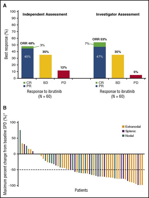
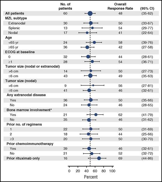
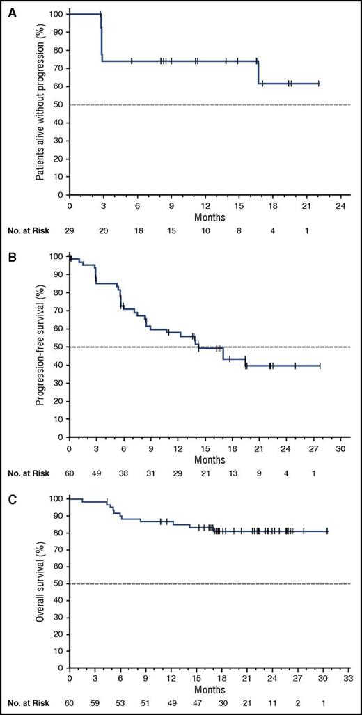
Three patients had nonmeasurable disease at baseline by IRC evaluation and were excluded from the efficacy analysis; thus, 60 patients were evaluable for efficacy. By independent assessment, ORR was 48% (95% CI, 35-62) with CR in 2 patients (3%). In a sensitivity analysis, ORR by investigator assessment was 53% (95% CI, 40-66) with CR in 4 patients (7%) (Figure 1A). The concordance rate for ORR between independent and investigator assessments was 84%. With clinical benefit defined as stable disease or better, 83% had clinical benefit by independent assessment and 88% by investigator assessment. Tumor reduction was noted in 78% (49 of 63 all-treated patients) by investigator assessment (Figure 1B). Median time to initial response was 4.5 months (range, 2.3-16.4), and median time to best response was 5.2 months (range, 2.3-16.4). ORR appeared consistent across disease characteristics (Figure 2). Details of responses by MZL subtypes are shown in the supplemental Appendix (see supplemental Table 2 for the evaluable population and supplemental Table 3 for the intent-to-treat population). Median DOR was not reached (95% CI, 16.7 to not estimable [NE]) per independent assessment (Figure 3A), with 62% of responders alive and progression-free at 18 months; median DOR was not reached in any of the subgroups by MZL subtype. By investigator assessment, median DOR was 19.4 months (95% CI, 7.3 to NE). Treatment-related lymphocytosis (defined as increase in absolute lymphocyte count of ≥ 50% from baseline to ≥5000/µL) was reported in 7 patients (11%; 1 patient had only a single elevated laboratory value); median time to peak absolute lymphocyte count was 1 week (range, 1-24), and median time to resolution was 11 weeks (range, 5-29).
ORRs and waterfall plot for reductions in lymph node sum of the products of longest diameters (SPD). (A) ORRs as assessed by the independent review committee and by investigators. (B) Waterfall plot for maximum percentage decrease in lymph node SPD by investigator assessment. *Data based on investigator assessment for 59 patients; 4 patients who discontinued treatment prior to first response assessment were not evaluable. Investigator-assessed data were used given that IRC assessment included 2 separate sets of SPD data due to readings by 2 radiologists.
ORRs and waterfall plot for reductions in lymph node sum of the products of longest diameters (SPD). (A) ORRs as assessed by the independent review committee and by investigators. (B) Waterfall plot for maximum percentage decrease in lymph node SPD by investigator assessment. *Data based on investigator assessment for 59 patients; 4 patients who discontinued treatment prior to first response assessment were not evaluable. Investigator-assessed data were used given that IRC assessment included 2 separate sets of SPD data due to readings by 2 radiologists.
ORRs according to subgroup. Forest plot representing subgroup analyses of ORRs as assessed by the independent review committee in the efficacy population (N = 60). The sizes of the circles are proportional to the sizes of the subgroups; error bars indicate 95% CIs. The dashed vertical line represents the overall treatment effect for all patients. *Results of bone marrow assessment were considered indeterminate in 4 patients.
ORRs according to subgroup. Forest plot representing subgroup analyses of ORRs as assessed by the independent review committee in the efficacy population (N = 60). The sizes of the circles are proportional to the sizes of the subgroups; error bars indicate 95% CIs. The dashed vertical line represents the overall treatment effect for all patients. *Results of bone marrow assessment were considered indeterminate in 4 patients.
DOR, PFS, and OS with ibrutinib. (A) DOR as assessed by the IRC. (B) PFS in the efficacy population (N = 60) as assessed by the IRC. (C) OS in the efficacy population (N = 60). The median PFS was 14.2 months (95% CI, 8.3 to NE), and the median OS has not been reached. The tick marks indicate patients with censored data.
DOR, PFS, and OS with ibrutinib. (A) DOR as assessed by the IRC. (B) PFS in the efficacy population (N = 60) as assessed by the IRC. (C) OS in the efficacy population (N = 60). The median PFS was 14.2 months (95% CI, 8.3 to NE), and the median OS has not been reached. The tick marks indicate patients with censored data.
With a median follow-up of 19.4 months (95% CI, 17.6-22.3) in the efficacy population, median PFS was 14.2 months (95% CI, 8.3 to NE) by independent assessment (Figure 3B) and 15.0 months (95% CI, 12.0 to NE) by investigator assessment. Median PFS by MZL subtype was 13.8 months (95% CI, 8.3 to NE) for extranodal, 19.4 months (95% CI, 8.2 to NE) for splenic, and 8.3 months (95% CI, 2.8 to NE) for nodal MZL. Median OS has not been reached (Figure 3C) with an 18-month estimated OS rate of 81% (95% CI, 68-89). Pseudoprogression occurred in 2 patients at weeks 9 and 13, respectively, confirmed by further follow-up (supplemental Appendix 2, “Patient cases of pseudoprogression”).
Safety
All 63 patients were evaluated for safety. The most common treatment-emergent AEs (any grade occurring in ≥20% of patients) are shown in Table 2. Treatment-emergent grade ≥3 AEs occurred in 42 patients (67%), with anemia (14%), pneumonia (8%), and fatigue (6%) among the most common events (Table 2). Grade ≥3 infections occurred in 12 patients (19%) overall, including a grade 5 infection (parainfluenza virus infection) in a patient with multiple system organ failure. Treatment-emergent serious AEs of any grade occurred in 28 patients (44%), with grade 3 or 4 pneumonia being the most common event reported in 5 patients (8%) (Table 2; full summary in supplemental Table 4). Bleeding AEs occurred in 37 patients (59%; full summary in supplemental Table 5), which were all grade 1-2 events except for 1 patient (2%) with major bleeding resulting in death (grade 5 cerebral hemorrhage, described below; the patient received anticoagulation with dalteparin). Twenty-nine patients (46%) received concomitant anticoagulants (16%) or antiplatelet agents (37%) during the study; aspirin (24%) was the most common antiplatelet agent used. Atrial fibrillation occurred in 4 patients (6%), which were all grade 1-2 events and did not lead to dose reduction or treatment discontinuation. All 4 patients had a prior history of known risk factors, including hypertension, diabetes mellitus, hypothyroidism, congestive heart failure, and/or coronary artery disease. AEs leading to dose reductions occurred in 6 patients (10%), with fatigue (3%) and stomatitis (3%) being the most common; 4 patients had 1 dose reduction, and 2 patients had 2 dose reductions. Diarrhea was the most common AE leading to treatment discontinuation, occurring in 3%. The full list of AEs leading to treatment discontinuation is included in supplemental Table 6.
Summary of adverse events
| Event . | Total (N = 63) . |
|---|---|
| Most common adverse events of any grade* | |
| Fatigue | 28 (44) |
| Diarrhea | 27 (43) |
| Anemia | 21 (33) |
| Nausea | 16 (25) |
| Arthralgia | 15 (24) |
| Peripheral edema | 15 (24) |
| Thrombocytopenia | 15 (24) |
| Cough | 14 (22) |
| Dyspnea | 13 (21) |
| Upper respiratory tract infection | 13 (21) |
| AEs grade ≥3† | |
| Anemia | 9 (14) |
| Pneumonia | 5 (8) |
| Fatigue | 4 (6) |
| Cellulitis | 3 (5) |
| Diarrhea | 3 (5) |
| Hypertension | 3 (5) |
| Lymphocyte count decreased | 3 (5) |
| Neutropenia | 3 (5) |
| Asthenia | 2 (3) |
| Autoimmune hemolytic anemia | 2 (3) |
| Blood bilirubin increased | 2 (3) |
| Muscle spasms | 2 (3) |
| Multiple organ dysfunction | 2 (3) |
| Neutrophil count decreased | 2 (3) |
| Pneumothorax | 2 (3) |
| Sepsis | 2 (3) |
| Serious AEs‡ | |
| Pneumonia | 5 (8) |
| Cellulitis | 2 (3) |
| Autoimmune hemolytic anemia | 2 (3) |
| Pneumothorax | 2 (3) |
| Sepsis | 2 (3) |
Data are presented as n (%) of patients unless indicated otherwise.
The events listed are AEs of any grade that occurred in at least 20% of patients.
The events listed are AEs of grade 3 or higher that occurred in at least 2 patients (3%).
The events listed are serious AEs that occurred in at least 2 patients (3%). The list of all serious AEs is provided in supplemental Table 3.
Three treatment-emergent AEs resulting in death were reported. These AEs included progressive disease (after 30 days of treatment), cerebral hemorrhage (19 days after discontinuing ibrutinib, and reported as unlikely related to study treatment, as the patient received anticoagulation with dalteparin prior to the bleeding event), and multiple system organ failure (in a patient with extensive pulmonary involvement by lymphoma who contracted parainfluenza pneumonia). Eight patients died during the study. Their deaths were attributable to progressive disease (4 patients), intracranial hemorrhage (1 patient; 57 days after discontinuing ibrutinib, which was outside the AE reporting period and considered secondary to anticoagulation for deep vein thrombosis), or unknown reasons (3 patients; 114, 304, and 347 days after discontinuing ibrutinib, which was outside the AE reporting period).
Discussion
Treatment of advanced or relapsed MZL is commonly approached with regimens used for other indolent NHLs such as follicular lymphoma.7 However, treatment-related toxicities may limit the options, particularly in an older MZL population (median age of diagnosis: 68 years).25 Moreover, the clinical course of MZL is heterogeneous, with nodal MZL (particularly for disseminated disease) associated with less favorable prognosis than other subtypes.2,9,25 Unfortunately, patients with MZL are often underrepresented or excluded from studies of other more common indolent NHLs. Our study represents one of the largest prospective trials conducted in all subtypes of MZL designed to identify an active single agent with a favorable toxicity profile.
In this study, single-agent ibrutinib demonstrated an ORR of 48% by independent assessment in previously treated patients with MZL. Notably, ORR was consistent across baseline clinical parameters, including the subgroups with bulky disease, bone marrow involvement, or prior chemoimmunotherapy. ORR was similar between subgroups with and without prior chemoimmunotherapy (46% vs 52%). Patients without prior chemoimmunotherapy had received rituximab monotherapy (n = 16) or rituximab monotherapy and other targeted agents or chemotherapy combinations (n = 5; CPI-0610, bendamustine, chlorambucil, or cyclophosphamide/doxorubicin/vincristine). The rate of disease control based on clinical benefit was 83%. More importantly, 78% benefited with reductions in tumor burden, consistent with the notion that BTK inhibition has biological relevance in MZL. Responses in this pretreated population were durable in all MZL subtypes, with a median DOR that was not reached at a median follow-up of 19 months.
Earlier published studies in MZL largely involved treatment-naive patients in whom responses are expected to be higher and more durable than in the relapsed/refractory setting. In a phase 2 trial of rituximab monotherapy in 34 patients with extranodal MZL, the ORR was 73% (CR in 44%) with a median DOR of 10.5 months.6 ORR in the subgroup without prior chemotherapy (n = 23) was 87% (CR in 48%), with a median time to treatment failure of 22 months; the subgroup that received prior chemotherapy regimens, including CVP (cyclophosphamide, vincristine, and prednisone), cladribine, fludarabine, or anthracycline-containing combinations (n = 11) showed less favorable outcomes, with an ORR of 45% (CR in 36%) and a median time to treatment failure of 12 months. Rituximab in combination with chemotherapy (alkylating agents or purine analogs) has shown ORRs of 85% to 93% (CR in 54% to 78%) in predominantly chemotherapy-naive or treatment-naive patients.4,5,9,12,26
In the relapsed setting, a retrospective analysis of 26 patients with extranodal MZL (no prior systemic antitumor therapy in 10 patients; 38%) treated with rituximab combined with cyclophosphamide, doxorubicin/mitoxantrone, vincristine, and prednisolone (R-CHOP or R-CNOP) showed an ORR of 77% (CR in 23%) with 85% of patients in continuous remission after a median follow-up of 19 months.10 Grade 3-4 cytopenias developed in 31% of patients.10 In another retrospective study in 14 patients with relapsed extranodal MZL (no prior systemic antitumor therapy in 6 patients; 43%), bendamustine combined with rituximab (BR) showed an ORR of 93% (CR in 71%) and durable remissions, with 12 patients (86%) remaining in remission at a median follow-up of 23 months.27 Grade 3-4 cytopenias occurred in 29% of these patients.27 In the randomized StiL trial, 230 patients with various subtypes of relapsed indolent NHLs were treated with BR (MZL, n = 10) or fludarabine combined with rituximab (MZL, n = 8); BR relative to fludarabine plus rituximab resulted in higher ORR (82% vs 51%) and CR rates (40% vs 17%) with longer median PFS (34 vs 12 months).11 However, response rates specifically in the MZL populations were not reported. Although chemoimmunotherapy with R-CHOP (or R-CHOP-like) or BR is standard treatment of other indolent NHLs such as follicular lymphoma, data on the use of these regimens in patients with MZL are limited. The above studies included only a small number of patients with MZL or a mixed population with indolent lymphomas, and they included “relapsed” patients who had received only local therapy (eg, surgery or radiation therapy) or H pylori eradication treatment without prior systemic antitumor therapy. Moreover, grade 3-4 myelosuppression was reported in a large proportion of patients treated with these chemotherapy or chemoimmunotherapy regimens.
Our trial can best be viewed in the context of other single-agent treatments for relapsed/refractory indolent NHL. In rituximab-exposed patients with low-grade or follicular lymphoma, retreatment with rituximab resulted in an ORR of 40% (CR in 11%) and a median time to progression of 18 months.28 Bendamustine monotherapy in rituximab-refractory indolent NHL showed an ORR of 75% (CR in 14%) with median DOR of 9 months; of the 16 patients with MZL, ORR was 78% (CR in 11%) in patients with nodal MZL (n = 9) and 86% (CR in 43%) in extranodal MZL (n = 7).8 Bendamustine was associated with a high rate of grade 3-4 cytopenias (neutropenia in 61%; thrombocytopenia in 25%); grade 3-4 infections were reported in 21%.8
More recent studies have also evaluated single-agent treatment with other targeted agents or immunomodulating therapy in patients with MZL or other indolent NHL subtypes. Lenalidomide monotherapy in previously untreated and relapsed/refractory extranodal MZL (N = 18; gastric mucosa-associated lymphoid tissue [MALT], n = 5; extragastric, n = 13) was associated with an ORR of 61% (CR in 33%).29 Responses occurred in 3 of 7 patients (43%) with prior systemic therapy. Toxicities were mainly nonhematologic AEs, and grade 3-4 neutropenia was reported in 17%.29 Bortezomib monotherapy was evaluated in patients with extranodal MALT lymphoma who had relapsed or were refractory to chemotherapy, antibody therapy, or a combination of both (N = 32).30 The major primary sites of involvement were gastric MALT (n = 14), skin (n = 14), lung (n = 4), and other sites (n = 3). Of 29 evaluable patients, the ORR was 48% (CR in 31%) with median DOR that was not reached at a median follow-up of 24 months.30 Neurological AEs were common and occurred in 65% of patients (including grade 3-4 peripheral sensory neuropathy in 13%); other common grade 3-4 AEs included neutropenia, thrombocytopenia, and fatigue (in 13% each).30 More recently, in a phase 2 study of the PI3Kδ-inhibitor idelalisib in rituximab-refractory indolent NHL, the ORR was 57% (CR in 6%), with a median DOR of 12.5 months.31 In the small subgroup with MZL (n = 15), the ORR was 47% (CR in 7%) with a median DOR of 18 months and median PFS of 7 months.32 Although idelalisib was generally well tolerated, grade ≥3 diarrhea, colitis, or both occurred in 16% of patients.31
Treatment with single-agent ibrutinib in our study was well tolerated, with an AE profile consistent with that of published studies of ibrutinib in previously treated NHL and CLL.23,33-36 AEs were largely grade 1-2 events, and the discontinuation rate due to AEs was low. The rate of grade ≥3 infections (19%), with pneumonia being the most common infection (8%), was similar to the rates reported with ibrutinib in patients with relapsed/refractory MCL (25%; grade ≥3 pneumonia in 6%).35 Preliminary data with short term follow-up from the phase 2 Consortium Trial of ibrutinib in relapsed/refractory follicular lymphoma reported grade 3-4 infections in 5% of patients and grade 5 pneumonia in 3%.37 In terms of major hemorrhage, only 1 case was reported as an AE (occurring 19 days after ibrutinib discontinuation) despite 46% of patients receiving concomitant anticoagulation or antiplatelet therapy during the study. Although 44% of patients experienced a serious AE, dose delays and adjustments allowed many patients to remain on therapy (exposure ≥12 months in 48%; median relative dose intensity 98%), and 38% were continuing ibrutinib at the time of analysis.
The rates of grade ≥3 and serious AEs appear consistent with those reported with single-agent ibrutinib in previously treated patients with other types of B-cell malignancies. It should be noted, however, that the median treatment duration (11.6 months) and median follow-up (19.4 months) in our study was longer relative to those in previously published studies, making direct comparisons of AE data across studies difficult. In the ibrutinib arm of the phase 3 RESONATE (PCYC-1112) study in previously treated CLL (420 mg once daily with median treatment duration 8.6 months; median follow-up 9.4 months), the rates of grade 3-4 AEs and serious AEs were 51% and 42%, respectively.33 These rates in the chlorambucil arm of the study (with median treatment duration of 5.3 months) were 39% and 30%, respectively. Importantly, grade ≥3 infections occurred at a similar rate between treatment arms, with 24% in the ibrutinib arm and 22% in the chlorambucil arm.33 Of relevance, the AE rates reported above were not adjusted for differences in treatment exposure (median 8.6 vs 5.3 months between the arms). In the phase 2 Consortium Trial of ibrutinib in patients with relapsed/refractory follicular lymphoma (560 mg once daily with median treatment duration 5 months; median follow-up 6.5 months), grade 3-4 AEs were reported in 30% of patients.37 In the phase 2 trial of ibrutinib in relapsed/refractory MCL (560 mg once daily with median treatment duration 9 cycles; median follow-up 15.3 months), serious AEs were reported in 56% of patients.35 Results from the current study also appear to compare favorably to other regimens used for MZL and indolent NHL, where substantial hematologic and infectious toxicities may limit their use.4,8,10,11,38
In conclusion, single-agent ibrutinib showed a high ORR in patients with relapsed/refractory MZL, which was consistent across clinical characteristics. Further, responses to ibrutinib were durable across all MZL subtypes. Importantly, in targeting BTK, our study has demonstrated that BCR signaling is an essential pathway in this lymphoma subtype. Also, due to evidence of pseudoprogression in our trial, as well as in follicular lymphoma (FLR2002 trial),39 biopsies may be warranted to differentiate between true lymphoma progression and immune-mediated antitumor response potentially triggered by ibrutinib-mediated inhibition of interleukin-2–inducible T-cell kinase (ITK) or other Tec kinases.40 Ibrutinib, now indicated for patients with MZL who require systemic therapy and have received at least 1 prior anti-CD20–based therapy, represents a new treatment option with a favorable benefit-risk profile and convenient, once-daily oral administration. Future studies will investigate ibrutinib in treatment-naive patients or as combination strategies in relapsed/refractory MZL.
The online version of this article contains a data supplement.
The publication costs of this article were defrayed in part by page charge payment. Therefore, and solely to indicate this fact, this article is hereby marked “advertisement” in accordance with 18 USC section 1734.
Acknowledgments
The authors thank the patients participating in this trial and their supportive families. They also thank Lorinda Soma (Department of Laboratory Medicine, University of Washington), John Katsetos (Hofstra Northwell School of Medicine), and Silvat Sheikh-Fayyaz (Division of Flow Cytometry/Urovysion, Long Island Jewish Medical Center, and Department of Pathology and Laboratory Medicine, North Shore Long Island Jewish Health System) for their contributions to the pathology assessments in this study.
This study was funded by Pharmacyclics LLC, an AbbVie Company. G.P.C. acknowledges support from the Blood theme of the Oxford NIHR Biomedical Research Centre. Maoko Naganuma, CMPP, funded by Pharmacyclics LLC, an AbbVie Company, provided medical writing support in the preparation of this manuscript.
Authorship
Contribution: A.N., D.M.B., and B.M. designed the study; A.N., S.d.V., C.T., P.M., C.R.F., F.M., G.P.C., S.M., M.C., S.P., S.S., J.C.B., and R.C. enrolled patients and collected data; B.M. performed statistical analysis of data; B.M., D.M.B., I.D., and A.S. assembled the data; B.M., D.M.B., I.D, and A.N. analyzed and interpreted the data; A.N. wrote the first draft of the manuscript; all authors were involved in the preparation of and revisions to the manuscript, and approved the final version of the manuscript for submission.
Conflict-of-interest disclosure: A.N. has received research funding from Pharmacyclics. C.T. has consulted for Janssen, Gilead, and Roche. P.M. has consulted for and received honoraria from Janssen and Celgene, consulted for Gilead, Acerta, and Novartis, and received research funding from Teva; he has also received travel accommodations from Janssen and Gilead. C.R.F. has consulted without compensation for Gilead and Genentech/Roche; he has also received research funding from Pharmacyclics, AbbVie, Acerta, Genentech/Roche, Gilead, Infinity, Millennium/Takeda, TG Therapeutics, ECOG, Mayo Clinic, and the National Institutes of Health. F.M. has served in an advisory role for and received honoraria from Roche, Celgene, Bristol-Meyers Squibb, and Janssen; he has also consulted for Servier and Gilead. G.P.C. has consulted for, received honoraria from, and served on a speakers’ bureau for Takeda, Roche, and Gilead; he has also consulted for and received honoraria from ADC Therapeutics and Bristol-Myers Squibb, received research funding from Celgene and Amgen, and received travel accommodations from Takeda and Napp. S.M. has consulted for and has served on a speakers’ bureau for Pharmacyclics/Janssen, Gilead, and Genentech; she has also received research funding from Pharmacyclics, Novartis, Xeme, Gilead, AbbVie, and Celgene. M.C. has held a leadership role in Immunomedics and holds stock ownership in Immunomedics and AbbVie; he has received research funding and has served on a speakers’ bureau for Pharmacyclics, Gilead, and Celgene, has consulted for Gilead and Celgene, and received research funding from Pfizer, GlaxoSmithKline, and Karyopharm. S.P. has received honoraria from, served on a speakers’ bureau for, and received travel accommodations from Takeda, Novartis, Celgene, Onyx, and Seattle Genetics; he has also consulted for Takeda. S.S. has received research funding from Pharmacyclics, Janssen, Merck, Acerta, and Genentech. J.C.B. has consulted for and has received research funding from AbbVie and Gilead and has consulted for Janssen. A.S., B.M., I.D., and D.M.B. are employed by Pharmacyclics LLC, an AbbVie Company, and hold stock ownership in AbbVie. R.C. has consulted for and has served in a speakers’ bureau for Seattle Genetics, Millennium, and Genentech; he has also received research funding from Pharmacyclics, Seattle Genetics, Millennium, and Merck. S.d.V. declares no competing financial interests.
Correspondence: Ariela Noy, Department of Medicine, Lymphoma Service, Memorial Sloan Kettering Cancer Center, 1275 York Ave, New York, NY 10021; e-mail: noya@mskcc.org.


