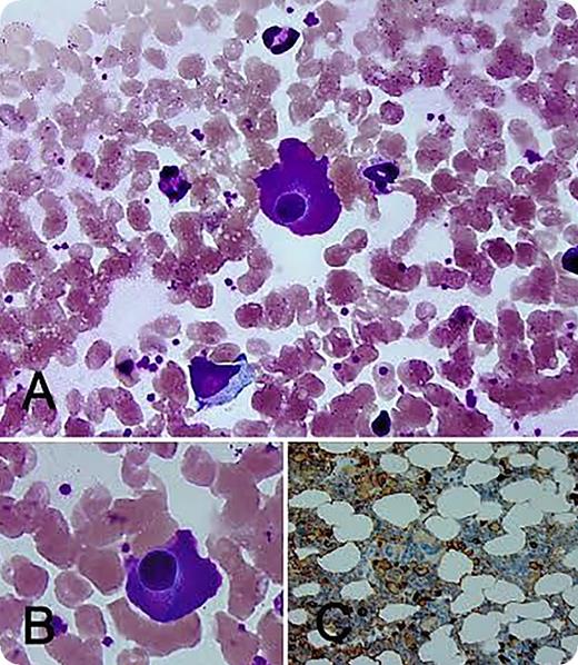
A 56-year-old diabetic woman presented with significant proteinuria. Serum protein electrophoresis demonstrated an elevated α2 region. Immunofixation confirmed immunoglobulin A-λ (IgA-λ) monoclonal protein (unquantifiable). The serum IgA level was 763 mg/dL (normal range, 83-407 mg/dL). The serum κ/λ free light chain ratio was 0.5 (normal range, 0.26-1.76). The result of urine protein electrophoresis was negative for monoclonal proteins. Blood counts, blood chemistry, and skeletal survey findings were normal. The bone marrow aspirate smear showed plasma cells with abundant eosinophilic cytoplasm consistent with flame cells (panels A-B; original magnification ×100; Wright-Giemsa stain). There was a λ light chain (dim) restricted plasma cell population (∼5% on bone marrow core biopsy specimen) (panel C; original maginification ×40; CD138 immmunohistochemistry stain). Overall, findings were consistent with IgA monoclonal gammopathy of undetermined significance.
Historically, flame cells were used to identify IgA monoclonal proteins and are more often reported in association with IgA myeloma. The “flaming” phenomenon can be uncommonly seen in other myeloma types and in reactive inflammatory states. There is no definitive evidence to suggest an association with myeloma progression or aggressiveness. The deeply stained pinkish hue at the cell periphery is thought to be the result of precipitated carbohydrate-rich IgA in the endoplasmic reticulum, which appears in stark contrast to the perinuclear halo of the Golgi apparatus. The patient continues to receive follow-up care and has stable renal function, which is now presumed to be diabetes related.
For additional images, visit the ASH IMAGE BANK, a reference and teaching tool that is continually updated with new atlas and case study images. For more information visit http://imagebank.hematology.org.


This feature is available to Subscribers Only
Sign In or Create an Account Close Modal