Abstract
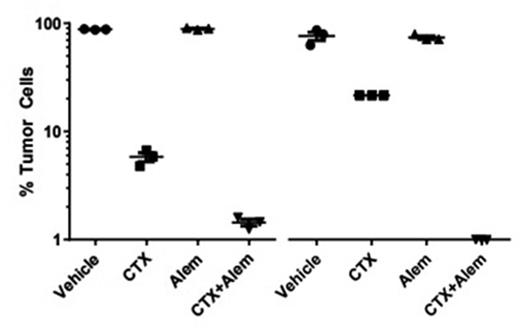
Double Hit Lymphomas (DHL) constitute 6-9% of all newly-diagnosed Diffuse Large B-cell Lymphomas (DLBCL) and are defined by MYC translocation in concert with BCL2 (80%) or BCL6 (20%) rearrangement. DHL is characterized by poor upfront response to standard chemoimmunotherapies. The development of more effective treatment for DHL has been limited by the lack of models that accurately recapitulate the biology of this disease. To faithfully model bona fide human DHL, we generated two patient-derived xenografts (PDXs) in NSG mice from patients with DHL who relapsed after or were refractory to R-CHOP. Both PDXs had low/negative expression of surface CD20, in line with previous reports of DHL, and high surface expression of CD52. Importantly, these features recapitulated the original tumor phenotypes. We reasoned that immunologic approaches that target DHLs may help overcome their resistance to cytotoxic chemotherapy. Given the surface expression of CD52 on both PDXs, we treated xenografted mice with the anti-CD52 antibody Alemtuzumab (2x5mg/kg) when >2% tumor was detected in peripheral blood, a time corresponding to marked splenomegaly and ~30% bone marrow involvement. In both models, Alemtuzumab markedly reduced tumor involvement in the peripheral blood and spleen; however, the bone marrow was largely refractory to Alemtuzumab treatment. This is in line with previous results (Pallasch et al. Cell 2014) who also observed bone marrow-specific resistance to Alemtuzumab using a humanized transgenic model of DHL. In the latter model, Alemtuzumab resistance was successfully overcome by high-dose cyclophosphamide (CTX, 100mg/kg), which induced a tumor secretory response that induced macrophage-mediated phagocytosis of tumor cells. Thus, we treated both PDX models with vehicle, CTX 100mg/kg, Alemtuzumab or CTX+Alemtuzumab. While CTX monotherapy was able to partially reduce bone marrow tumor burden, in both PDXs, a single dose of CTX+Alemtuzumab was highly synergistic and eradicated nearly all bone marrow disease (Figure 1), which resulted in significantly extend murine survival compared to either CTX or Alemtuzumab alone alone (p<0.01). In contrast, high-dose Doxorubicin (DOX, 5mg/kg) had minimal effect on bone marrow DHL and DOX+Alemtuzumab was not synergistic. Strikingly, CD11b+F4/80+CD11c- macrophages comprised 20-40% of bone marrow cellularity 8 days after CTX treatment of xengorafted mice. DHL cells isolated from treated mice were next examined for changes in surface protein expression. At 16 hours after treatment, bone marrow DHL cells in mice receiving CTX (but not those receiving DOX or vehicle) had significantly reduced levels of surface CD47, the “don't eat me” signal that suppresses macrophage phagocytosis. By 48 hours after treatment, bone marrow DHL cells in mice receiving CTX (but not those receiving DOX or vehicle) had significantly increased expression of the pro-phagocytic proteins CD31 and Calreticulin. Next, we performed cytokine array analysis on bone marrow from mice engrafted with DHL PDXs at 16 hours after treatment. CTX-treated (but not vehicle- or DOX-treated) bone marrow had significantly increased levels of human IL-16 and CCL4, cytokines known to promote monocyte recruitment, as well as VEGF, PDGF and TNFα. Ex-vivo phagocytosis assays using murine bone marrow-derived macrophages confirmed that recombinant human VEGF and TNFα each enhanced macrophage phagocytosis of Alemtuzumab coated Raji lymphoma cells. Findings from RNA-seq studies on tumor cells and macrophages from the spleen and bone marrow as well as therapy induced changes in these respective niches will be further discussed at the meeting. In summary, we have demonstrated that high-dose CTX induces a phenotypic and secretory response from human DHL cells in vivo that promotes phagocytosis of Alemtuzumab-opsinized cells. As a result, we are initiating a clinical trial of this combination in patients with relapsed/refractory DHL.
Percent tumor cells isolated from bone marrow of mice xenografted with DHL #61614 (left) or #63014 (right) on day 8 after treatment with cyclophosphamide (CTX), alemtuzumab (Alem) or the combination. Each marker indicates a mouse.
Percent tumor cells isolated from bone marrow of mice xenografted with DHL #61614 (left) or #63014 (right) on day 8 after treatment with cyclophosphamide (CTX), alemtuzumab (Alem) or the combination. Each marker indicates a mouse.
No relevant conflicts of interest to declare.
Author notes
Asterisk with author names denotes non-ASH members.


This feature is available to Subscribers Only
Sign In or Create an Account Close Modal