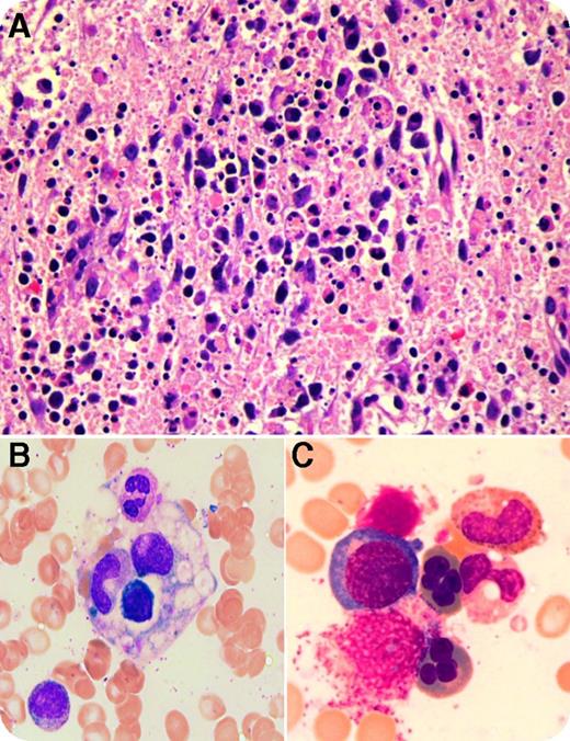
A 37-year-old man of Pakistani origin presented with a 5-week history of fever, night sweats, fatigue, nausea, vomiting, lymphadenopathy, and splenomegaly. He had similar symptoms 5 years prior with a cervical lymph node (LN) biopsy showing histiocytic necrotizing lymphadenitis or Kikuchi disease (KD). He had pancytopenia, with a neutrophil count of 0.52 × 10⁹/L, hemoglobin count of 76 g/L, mean corpuscular volume of 82.2, platelet count of 96 × 109/L, elevated erythrocyte sedimentation rate of 51, lactate dehydrogenase of 531 U/L, ferritin of 10 153 µg/L, triglycerides of 4.12 mmol/L, alanine transaminase of 277 U/L, aspartate transaminase of 418 U/L, and γ-glutamyl transferase of 215 U/L. Repeat LN biopsy suggested KD with preserved architecture and patchy fibrinoid necrotizing foci with numerous apoptotic bodies and cellular debris admixed with lymphocytes and histiocytes (panel A; original magnification ×40; hematoxylin and eosin stain). Extensive microbiological, rheumatologic, and hematological investigations, including immunohistochemistry, flow cytometry, molecular tests, etc, were noncontributory but ruled out lymphoma and the acute form of lupus. Marrow was reactive with increased histiocytes showing hemophagocytosis (panel B; original magnification ×100, oil immersion; May-Grünwald Giemsa stain) and dyserythropoiesis including multinuclearity (panel C; original magnification ×100, oil immersion; May-Grünwald Giemsa stain). Cytogenetics was normal. A diagnosis of hemophagocytic lymphohistiocytosis (HLH) with association of KD and dyserythropoiesis was rendered as he fulfilled 6 of 8 diagnostic criteria. The patient was treated with prednisone with complete recovery. He was doing well over 1 year after initial diagnosis.
The association of HLH and KD has been reported in limited cases, as has the association of HLH and reactive dysplasia. The coexistence of HLH, KD, and dysplasia is extremely rare but is worth further investigations.
For additional images, visit the ASH IMAGE BANK, a reference and teaching tool that is continually updated with new atlas and case study images. For more information visit http://imagebank.hematology.org.


This feature is available to Subscribers Only
Sign In or Create an Account Close Modal