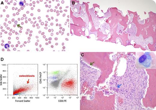
A 33-year-old African American man with end-stage renal disease on hemodialysis for 5 years presented with anemia and splenomegaly. His peripheral blood smear demonstrated anemia (hemoglobin 77 g/L) with anisopoikilocytosis including occasional teardrop cells (panel A, green arrow). Bone marrow (BM) core biopsy revealed a hypocellular marrow with trilineage hypoplasia (panel B), osteosclerosis with increased osteoblasts (panel C, green arrow/inset) and osteoclasts (blue arrow), and grade 3 myelofibrosis (with reticulin and collagen fibrosis). There was no evidence for a myeloproliferative neoplasm (MPN) or metastatic tumor. Cytogenetic/molecular studies for JAK2 V617F, MPL mutations, and BCR/ABL1 rearrangement were normal. Flow cytometric analysis (FCA) on the BM biopsy revealed CD56+/CD45− osteoblasts (panel D, red arrow; events in blue and green represent hematogones and T cells, respectively). The parathyroid hormone level was significantly increased (3295 pg/mL; reference range, 15-65 pg/mL). The overall findings were diagnostic of nonneoplastic myelofibrosis resulting from secondary hyperparathyroidism in a patient with long-standing end-stage renal disease.
Myelofibrosis secondary to renal osteodystrophy is an uncommon disease and has been rarely reported. Recognition of its histologic features is important in the differential diagnosis of a fibrotic BM (in particular, MPNs). Furthermore, the presence of CD56+/CD45− cells by FCA may be mistaken for evidence of a metastatic tumor. Comprehensive analysis is instrumental for an accurate diagnosis.
For additional images, visit the ASH IMAGE BANK, a reference and teaching tool that is continually updated with new atlas and case study images. For more information visit http://imagebank.hematology.org.


This feature is available to Subscribers Only
Sign In or Create an Account Close Modal