Key Points
Klf5 functions in hematopoiesis to regulate HSC and progenitor proliferation and localization in the bone marrow.
Klf5 is required in the granulocyte lineage and positively affects neutrophil output at the expense of eosinophil production.
Abstract
Krüppel-like factor 5 (Klf5) encodes a zinc-finger transcription factor and has been reported to be a direct target of C/EBPα, a master transcription factor critical for formation of granulocyte-macrophage progenitors (GMP) and leukemic GMP. Using an in vivo hematopoietic-specific gene ablation model, we demonstrate that loss of Klf5 function leads to a progressive increase in peripheral white blood cells, associated with increasing splenomegaly. Long-term hematopoietic stem cells (HSCs), short-term HSCs (ST-HSCs), and multipotent progenitors (MPPs) were all significantly reduced in Klf5Δ/Δ mice, and knockdown of KLF5 in human CD34+ cells suppressed colony-forming potential. ST-HSCs, MPPs, and total numbers of committed progenitors were increased in the spleen of Klf5Δ/Δ mice, and reduced β1- and β2-integrin expression on hematopoietic progenitors suggests that increased splenic hematopoiesis results from increased stem and progenitor mobilization. Klf5Δ/Δ mice show a significant reduction in the fraction of Gr1+Mac1+ cells (neutrophils) in peripheral blood and bone marrow and increased frequency of eosinophils in the peripheral blood, bone marrow, and lung. Thus, these studies demonstrate dual functions of Klf5 in regulating hematopoietic stem and progenitor proliferation and localization in the bone marrow, as well as lineage choice after GMP, promoting increased neutrophil output at the expense of eosinophil production.
Introduction
Krüppel-like-factor 5 (KLF5) is a member of the Kruppel-like family of transcription factors, several of which have been shown to regulate hematopoietic stem and progenitor cell function and lineage fate.1-4 Klf5 is a master regulator of growth, differentiation, migration, and apoptosis, is implicated as both a tumor suppressor and oncogene in the pathogenesis of many cancers, and is increasingly of interest as a therapeutic target.5,6 Previous in vitro studies have indicated that Klf5 plays a role in regulating myeloid growth and differentiation, particularly in the neutrophil lineage.7,8 Hypermethylation of KLF5 intron 1 occurs in 41% of acute myeloid leukemia (AML) patients, is an independent predictor of poor overall survival,9 and is associated with reduced expression of KLF5 mRNA.7,8 Thus, KLF5 may act as a tumor suppressor in AML, and further investigation of KLF5 function in the myeloid compartment is important for the understanding of this tumor suppressor activity. The KLF5 promoter is a direct binding target of C/EBPα,10,11 which is a master regulator of myelopoiesis, being essential for the transition of common myeloid progenitors (CMP) to granulocyte-macrophage progenitors (GMP).10,12 KLF5 gene expression is directly regulated by C/EBPα,10 consistent with reduced KLF5 expression in the CEBPA-mutated AML subgroup.13,14 Most recently, experiments in murine models have shown that C/EBPα-induced GMP formation is an essential step in leukemic transformation by the MLL-AF9 and MOZ-TIF2 oncogenes.12 Thus, we used the Cre-Lox system to generate mice with targeted ablation of Klf5 in the hematopoietic compartment. A detailed analysis of myeloid progenitor and mature cell compartments in these mice revealed that, although Klf5 is not absolutely required for CMP or GMP formation, it is important for neutrophil and eosinophil production, potentially functioning as a downstream target of C/EBPα after GMP formation to control lineage fate of myelomonocytic precursors.
Study design
Mice
Details for the mice used in this study are described in the supplemental Materials, available on the Blood Web site.
Flow cytometry analysis
Colony-forming unit assay
Methylcellulose assays for colony-forming cells (CFCs)17 were performed using MethoCult GF M3434 (mouse) or MethoCult H4034 Optimum (human) according to the manufacturer’s protocol.
For detailed methods regarding immunofluorescence, short hairpin RNA (shRNA) knockdown, and KLF5 overexpression, see supplemental Materials.
Results and discussion
To investigate the impact of Klf5 loss in hematopoiesis, we generated a conditional Klf5 gene ablation model (Klf5Δ/Δ mice) using the well-characterized Vav-cre transgene,18 to induce loss of Klf5 exon 2 in the hematopoietic system, thus removing the majority of the protein coding potential (supplemental Figure 1A). Quantitative polymerase chain reaction and western blot analysis indicated that Klf5 mRNA and protein were significantly reduced in whole bone marrow cells and CD45+ cells, respectively (supplemental Figure 1B-C). Klf5Δ/Δ mice were viable and born in expected Mendelian ratios with no observed effect on mortality in the time monitored (age, to 18 months; data not shown). However, longitudinal analysis identified a blood phenotype, characterized by a progressive increase in peripheral white blood cells and associated with increasing splenomegaly over time (supplemental Figure 2A-B).
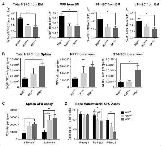
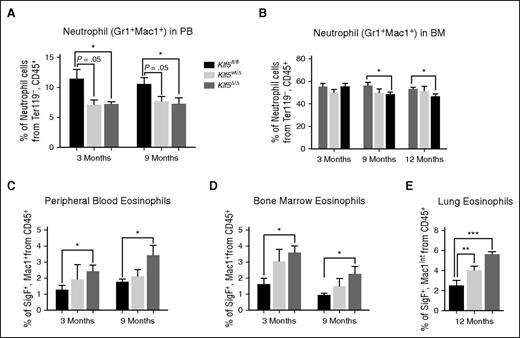
As C/EBPα acts as a master transcription factor early in adult hematopoiesis to regulate myelomonocytic cell output via GMP production, we next used flow cytometry to analyze the hematopoietic stem and progenitor cell (HSPC) fractions from bone marrow of Klf5Δ/Δ mice, using Klf5fl/fl littermates as controls. The fraction of long-term hematopoietic stem cells (LT-HSCs), short-term HSCs (ST-HSCs), and multipotent progenitors (MPPs) were all significantly reduced in Klf5Δ/Δ mice (Figure 1A), consistent with high relative expression of KLF5 in both mouse and human HSC compartments (supplemental Figure 3) and the previously reported function of Klf5 in hematopoietic stem cells.19 As Ishikawa et al19 showed that Klf5 regulates homing and adhesion of HSCs in the bone marrow, we hypothesized that the reduction of HSCs in the bone marrow may be associated with splenic myelopoiesis due to increased mobility of Klf5Δ/Δ HSCs. As shown in Figure 1B, flow cytometric analysis revealed that HSPCs (predominantly the ST-HSCs and MPPs) were increased in the spleen of Klf5Δ/Δ mice as a fraction of total Lin−c-Kit+ cells. The total number of committed progenitors, as determined from clonal assays, was also increased in the spleen of Klf5Δ/Δ mice (Figure 1C). Reduced levels of β1- and β2-integrin proteins in c-Kit+ cells from bone marrow were observed (supplemental Figure 4), which also correlated with decreased mRNA in total bone marrow from Klf5Δ/Δ mice. Consistent with previous studies of hematopoietic stem and progenitor cells from Klf5-deficient mice,19 Rab5b, which is an upstream regulator of the β-integrins and a direct Klf5 target, also had decreased mRNA in total bone marrow from Klf5Δ/Δ mice (supplemental Figure 4). Therefore, it is likely that that reduced adhesion to bone marrow stroma contributes to increased HSC mobilization, resulting in the increased stem and progenitor numbers observed in the spleen. We also observed a change in replating capacity for Klf5Δ/Δ committed progenitors. Clonal serial replating assays using bone marrow mononuclear cells demonstrated a reduction in colony numbers for Klf5Δ/Δ cells following secondary and tertiary plating (Figure 1D). This is consistent with reduced self-renewal capacity associated with loss of Klf5 function, and this defect may be inherited from the HSCs as a previous report found that the repopulation ability of the long-term HSC compartment was impaired in Klf5-deficient mice.19 To investigate the effect of reduced KLF5 in human stem and progenitor cells, we targeted KLF5 for knockdown in CD34+ bone marrow cells. As shown in supplemental Figure 5, lentiviral-mediated targeting of KLF5 with 2 independent validated shRNA triggers reduced total CFCs, consistent with KLF5 having a conserved role in proliferation and survival of both mouse and human primitive hematopoietic cells. Examination of more committed progenitors in mouse bone marrow did not identify significant differences in the relative fraction of GMP or pre-GMP as a percentage of Lin−c-Kit+ cells (supplemental Figure 6). Thus, although KLF5 is a known target of C/EBPα, it is not essential for C/EBPα-induced GMP production in the mouse. We next investigated changes in the neutrophil, eosinophil, T-cell, and B-cell fractions in total leukocytes from peripheral blood and bone marrow of adult Klf5Δ/Δ mice. A significant reduction in the fraction of Gr1+Mac1+ cells (neutrophils) as early as 3 months of age in peripheral blood was observed, and this was still evident in bone marrow until ≥12 months of age (Figure 2A-B). Analysis of eosinophil levels using flow cytometry revealed an increase in the eosinophil fraction of total leukocytes in the peripheral blood, bone marrow, and lungs of Klf5Δ/Δ mice (Figure 2C,D,E, respectively), suggesting that Klf5 may function as a negative regulator of adult eosinophil development. Changes to other compartments in Klf5Δ/Δ mice, included an increase in the fraction of T cells (CD3+ cells) in the peripheral blood, but not in the bone marrow or spleen, and transient differences in the fraction of B cells (B220+ cells) in peripheral blood, bone marrow, and spleen (supplemental Figure 7). Flow cytometric analysis of erythroid and megakaryocyte progenitors also did not identify significant differences in the Klf5Δ/Δ mice (supplemental Figure 8).15,20
Measurement and functional characterization of HSPCs in Klf5Δ/Δ mice. (A) Measurement of total HSPC (defined by IL7Rα−c-Kit+Sca-1+), LT-HSCs (defined by c-kit+Lin−IL7Rα−Sca-1+CD48−CD150+), ST-HSCs (defined by c-kit+Lin−IL7Rα−Sca-1+CD48+CD150+), and MPPs (defined by c-kit+Lin−IL7Rα−Sca-1+CD48+CD150−) as a percentage of the cKit+Lin− compartment from bone marrow of mice at 9 months of age using flow cytometry. Results were obtained from n = 8 Klf5fl/fl, n = 8 Klf5wt/Δ, and n = 11 Klf5Δ/Δ mice. (B) Total HSPCs, MPPs, and ST-HSCs (as a percentage of the cKit+Lin− compartment) in the spleen of 12-month-old mice. Results were obtained from n = 7 Klf5fl/fl, n = 5 Klf5wt/Δ, and n = 8 Klf5Δ/Δ mice. (C) Enumeration of myelo-erythroid colonies from spleen cells of Klf5Δ/Δ mice at 9 and 12 months old. Results were obtained from n = 6 Klf5fl/fl, n = 5 Klf5wt/Δ, and n = 7 Klf5Δ/Δ mice at 3 months and n = 6 Klf5fl/fl, n = 4 Klf5wt/Δ, and n = 6 Klf5Δ/Δ mice at 9 months. (D) Quantification of myelo-erythroid progenitor self-renewal from bone marrow using serial colony-forming assays at 9 months. Results were obtained from n = 4 Klf5fl/fl, n = 5 Klf5wt/Δ, and n = 5 Klf5Δ/Δ mice. Mean and SEM are shown for all measurements. P values were obtained from 2-tailed t tests conducted between each genotype group: ***P < .001, **P < .01, and *P < .05.
Measurement and functional characterization of HSPCs in Klf5Δ/Δ mice. (A) Measurement of total HSPC (defined by IL7Rα−c-Kit+Sca-1+), LT-HSCs (defined by c-kit+Lin−IL7Rα−Sca-1+CD48−CD150+), ST-HSCs (defined by c-kit+Lin−IL7Rα−Sca-1+CD48+CD150+), and MPPs (defined by c-kit+Lin−IL7Rα−Sca-1+CD48+CD150−) as a percentage of the cKit+Lin− compartment from bone marrow of mice at 9 months of age using flow cytometry. Results were obtained from n = 8 Klf5fl/fl, n = 8 Klf5wt/Δ, and n = 11 Klf5Δ/Δ mice. (B) Total HSPCs, MPPs, and ST-HSCs (as a percentage of the cKit+Lin− compartment) in the spleen of 12-month-old mice. Results were obtained from n = 7 Klf5fl/fl, n = 5 Klf5wt/Δ, and n = 8 Klf5Δ/Δ mice. (C) Enumeration of myelo-erythroid colonies from spleen cells of Klf5Δ/Δ mice at 9 and 12 months old. Results were obtained from n = 6 Klf5fl/fl, n = 5 Klf5wt/Δ, and n = 7 Klf5Δ/Δ mice at 3 months and n = 6 Klf5fl/fl, n = 4 Klf5wt/Δ, and n = 6 Klf5Δ/Δ mice at 9 months. (D) Quantification of myelo-erythroid progenitor self-renewal from bone marrow using serial colony-forming assays at 9 months. Results were obtained from n = 4 Klf5fl/fl, n = 5 Klf5wt/Δ, and n = 5 Klf5Δ/Δ mice. Mean and SEM are shown for all measurements. P values were obtained from 2-tailed t tests conducted between each genotype group: ***P < .001, **P < .01, and *P < .05.
Characterization of mature myeloid cells in Klf5Δ/Δ mice using flow cytometry. Measurement of neutrophils (Gr1+Mac1+) from (A) peripheral blood and (B) bone marrow at 3 and 9 months. Eosinophils, measured as Mac1+SigF+ cells, from (C) peripheral blood, (D) bone marrow, and (E) lung (Mac1intSigF+). Results from peripheral blood and bone marrow were obtained from n = 10 Klf5fl/fl, n = 6 Klf5wt/Δ, and n = 10 Klf5Δ/Δ mice at 3 months and n = 8 Klf5fl/fl, n = 8 Klf5wt/Δ, and n = 10 Klf5Δ/Δ mice at 9 months. For lung eosinophil analysis, results were obtained from n = 6 Klf5fl/fl, n = 4 Klf5wt/Δ, and n = 6 Klf5Δ/Δ at 12 months. Mean and SEM are shown for all measurements. P values were obtained from 2-tailed t tests conducted between each genotype group: ***P < .001, **P < .01, or *P < .05.
Characterization of mature myeloid cells in Klf5Δ/Δ mice using flow cytometry. Measurement of neutrophils (Gr1+Mac1+) from (A) peripheral blood and (B) bone marrow at 3 and 9 months. Eosinophils, measured as Mac1+SigF+ cells, from (C) peripheral blood, (D) bone marrow, and (E) lung (Mac1intSigF+). Results from peripheral blood and bone marrow were obtained from n = 10 Klf5fl/fl, n = 6 Klf5wt/Δ, and n = 10 Klf5Δ/Δ mice at 3 months and n = 8 Klf5fl/fl, n = 8 Klf5wt/Δ, and n = 10 Klf5Δ/Δ mice at 9 months. For lung eosinophil analysis, results were obtained from n = 6 Klf5fl/fl, n = 4 Klf5wt/Δ, and n = 6 Klf5Δ/Δ at 12 months. Mean and SEM are shown for all measurements. P values were obtained from 2-tailed t tests conducted between each genotype group: ***P < .001, **P < .01, or *P < .05.
Thus, these studies, and those of Ishikawa et al19 in independent Klf5 murine gene ablation models, demonstrate that Klf5 functions in hematopoiesis to regulate HSC and progenitor mobilization and granulocyte-macrophage progenitor lineage choice after GMP, positively affecting neutrophil output at the expense of eosinophil production. Increased expression of KLF5 mRNA is observed in the granulocyte lineage during both mouse and human hematopoiesis7 (supplemental Figure 3), and KLF5 overexpression promotes differentiation in human U937 cells following retroviral transduction with increased granulocytes (evident morphologically and by CD11b expression), which is particularly observed following induction with ATRA (supplemental Figure 9). As we previously showed that KLF5 is silenced by methylation in U937 cells,7 this also has therapeutic relevance to hematopoietic disease states.
In addition to C/EBPα, other members of the CEBP family may also be important as Klf5 interacts directly with C/EBPβ,21 which has been reported as a regulator of eosinophil gene expression.22 Klf5 has also been reported as a C/EBPβ target gene,21 and therefore Klf5 may interact with, and function downstream of, a range of other hematopoietic transcription factors involved in specification of the granulocyte-macrophage lineage. For example, GFI1 and KLF4 have been shown to bind the KLF5 promoter.11 Interestingly, KLF4 and KLF5 are often coexpressed and are known to coordinately or antagonistically regulate cellular differentiation in a range of contexts.5,23 KLF4 controls monocyte lineage fate and is also a target of gene silencing in AML.3 Other KLFs also have roles in hematopoiesis and leukemia; for example, KLF1 is a master regulator of erythropoiesis and variation at the KLF1 locus underlies a large spectrum of red blood cell disorders.2 KLF7 regulates function of primitive hematopoietic cells,4 and KLF2 has been reported to function in lymphoid development and disease and in regulating adhesion of myeloid cells.24 Therefore, further work to dissect the functional relationships of different members of the KLF family in normal and malignant hematopoiesis will be important. Future experiments to define both the direct upstream regulators and downstream targets of KLF5 in the hematopoietic compartment will add to our knowledge of stem cell regulation and myeloid differentiation. Finally, reduced KLF5 activity associated with hypermethylation of KLF5 intron 1 may contribute to the mobilization of preleukemic or leukemic stem and progenitor cells, thus separating these cells from critical regulators of growth and differentiation.
The online version of this article contains a data supplement.
The publication costs of this article were defrayed in part by page charge payment. Therefore, and solely to indicate this fact, this article is hereby marked “advertisement” in accordance with 18 USC section 1734.
Acknowledgments
The authors thank Warren Alexander (Walter and Eliza Hall Institute of Medical Research) for the kind gift of Vav-cre transgenic mice; Detmold Family Cytometry Facility’s staff members for help with flow cytometry; Maung Kyaw Ze Ya for assistance with immunofluorescence; and SA Pathology animal care facility staff members for mouse husbandry services.
The study was supported financially by the National Health and Medical Research Council of Australia.
Authorship
Contribution: N.H.S. contributed to research design, performed the research, analyzed the data, and drafted the manuscript; S.D. contributed to research design, performed research, and analyzed the data; L.A.D. contributed to research design and critically reviewed the manuscript; and A.L.B. and R.J.D. contributed to research design, supervised the research, and critically reviewed and wrote the manuscript.
Conflict-of-interest disclosure: N.H.S. has received scholarship funding from Adelaide Graduate Research Scholarship.
Correspondence: Richard J. D’Andrea, Acute Leukaemia Laboratory, Division of Haematology, Centre for Cancer Biology, IMVS/SA Pathology, PO Box 14, Rundle Mall, Adelaide, SA 5000, Australia; e-mail: richard.dandrea@unisa.edu.au.
References
Author notes
A.L.B. and R.J.D. contributed equally to this work.


This feature is available to Subscribers Only
Sign In or Create an Account Close Modal