In this issue of Blood, Motley et al have identified a novel and unexpected mechanism for clearance of extravascular fibrin that is accomplished by a specific proinflammatory macrophage population and is dependent upon active plasmin, yet independent of known fibrinogen receptors.1
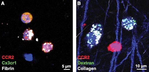
CCR2-positive cells endocytose fibrin, but not collagen. Cells positive for both CCR2 and CX3cr1 are identified by orange/yellow staining and show uptake of fibrin in white in panel A, whereas uptake of collagen, shown in white in panel B, is accomplished by cells that are negative for CCR2. Thus, cells that endocytose collagen are clearly distinct from cells that endocytose fibrin. See Figure 3H-I in the article by Motley et al that begins on page 1085.
CCR2-positive cells endocytose fibrin, but not collagen. Cells positive for both CCR2 and CX3cr1 are identified by orange/yellow staining and show uptake of fibrin in white in panel A, whereas uptake of collagen, shown in white in panel B, is accomplished by cells that are negative for CCR2. Thus, cells that endocytose collagen are clearly distinct from cells that endocytose fibrin. See Figure 3H-I in the article by Motley et al that begins on page 1085.
Fibrin is well known as the insoluble polymer that arrests bleeding. Fibrin formation is also essential in the extravascular space for formation of a scaffold during the initial phases of tissue remodeling during an inflammatory response. However, persistence of extravascular fibrin causes chronic inflammation and organ damage, and is a key feature in a wide variety of pathological processes including acute lung injury, asthma, idiopathic pulmonary fibrosis, rheumatoid arthritis, multiple sclerosis, autoimmune encephalitis, and cancer (reviewed in Schuliga2 and Bardehle et al3 ). The mechanism for initial extracellular degradation of fibrin by sequential proteolytic cleavages by the enzyme plasmin is well understood (plasmin is formed by activation of the circulating zymogen, plasminogen, by plasminogen activators, tissue-plasminogen activator [tPA] and urokinase [uPA]). However, the mechanisms by which fibrin degradation products are ultimately eliminated from the extravascular space remained an open area of investigation. Clues were provided by observations that leukocytes infiltrate extravascular fibrin deposits, and that cultured cells of the monocytoid lineage could endocytose fibrin. Here, Motley et al have identified a novel endocytic pathway used by macrophages that results in clearance of extravascular fibrin.1
Motley et al use a novel technique to quantify fibrin removal in vivo by implanting fluoresceinated fibrin films into the dermis of mice, from which specific molecules regulating fibrinolysis and/or inflammation were modulated or deleted. The fibrin films were removed and their weight and residual fluorescence quantified as a marker of fibrin degradation. In addition, the dermal implantation regions were subjected to intravital imaging to quantify the presence of fluoresceinated fibrin within lysosomes. Their results reveal that a distinct population of proinflammatory macrophages (that bear C-C chemokine receptor type 2 [CCR2] on their surface) is predominantly responsible for removal of fibrin by cellular uptake. Surprisingly, known fibrinogen receptors were not necessary for fibrin endocytosis, thus revealing the presence of a novel endocytotic pathway for fibrin removal. And the experimental models that influenced fibrin removal and cellular uptake pinpointed a requirement for active plasmin in this process. In other unanticipated results, the inflammatory macrophage population responsible for the cellular uptake of fibrin was clearly distinguished from the noninflammatory tissue-remodeling macrophage population responsible for uptake of collagen, as shown (see figure). The authors suggest that this makes sense in considering wound healing because fibrin is the initial matrix deposited when tissues are injured, which is later replaced by collagen during the tissue-remodeling phase.
The mechanism by which plasmin contributes to fibrin removal in vivo is intriguing. Clearly, initial extracellular production of fibrin degradation products by plasmin is necessary. In addition, plasmin activity promotes phagocytosis by both macrophages4 and dendritic cells.5 This involves both recognition of prey and direct effects on engulfment of prey, events which are regulated by plasmin-mediated activation of intracellular signaling pathways that regulate expression of major genes involved in phagocytosis.4 There may be an analogous mechanism for cellular fibrin uptake. It is also possible that plasminogen may bind to fibrin and bridge fibrin to the cell surface via cellular plasminogen receptors. Future studies to examine fibrin endocytosis in plasminogen receptor-deficient mice would be informative.6 In addition, mice deficient in thrombin-activatable fibrinolysis inhibitor (that removes plasmin[ogen] recognition sites from fibrin and from cell surfaces) may show enhanced fibrin uptake and could also shed light on these issues.
The results of Motley and colleagues provide a strong rationale for future studies to investigate the effect of deletion of CCR2-positive macrophages in disease models with a prominent extravascular fibrin deposition component. At the same time, the role of CCR2-positive macrophage endocytosis in intravascular fibrin clearance should be evaluated as this may have a profound effect on disease progression. For example, intravascular fibrin accumulation as well as extravascular fibrin accumulation are contributing features of Alzheimer disease7 and impaired recruitment of the CCR2-positive macrophage population is reported in Alzheimer disease patients (reviewed in Naert and Rivest8 ).
Thus, therapeutically increasing the number and/or activity of CCR2-positive macrophages may be an attractive therapeutic strategy for a variety of disease processes involving both extravascular and intravascular fibrin deposition.
Conflict-of-interest disclosure: The authors declare no competing financial interests.


This feature is available to Subscribers Only
Sign In or Create an Account Close Modal