Key Points
NE and EPI promote megakaryocyte adhesion, migration, and proplatelet formation via α2-adrenoceptor-ERK1/2 signaling.
Sympathetic stimulation enhances platelet production, which may facilitate recovery of thrombocytopenia or aggravate atherosclerosis.
Abstract
The effect of sympathetic stimulation on thrombopoiesis is not well understood. Here, we demonstrate that both continual noise and exhaustive exercise elevate peripheral platelet levels in normal and splenectomized mice, but not in dopamine β-hydroxylase–deficient (Dbh−/−) mice that lack norepinephrine (NE) and epinephrine (EPI). Further investigation demonstrates that sympathetic stimulation via NE or EPI injection markedly promotes platelet recovery in mice with thrombocytopenia induced by 6.0 Gy of total-body irradiation and in mice that received bone marrow transplants after 10.0 Gy of lethal irradiation. Unfavorably, sympathetic stress-stimulated thrombopoiesis may also contribute to the pathogenesis of atherosclerosis by increasing both the amount and activity of platelets in apolipoprotein E–deficient (ApoE−/−) mice. In vitro studies reveal that both NE and EPI promote megakaryocyte adhesion, migration, and proplatelet formation (PPF) in addition to the expansion of CD34+ cells, thereby facilitating platelet production. It is found that α2-adrenoceptor–mediated extracellular signal-regulated kinase 1/2 (ERK1/2) activation is involved in NE- and EPI-induced megakaryocyte adhesion and migration, and PPF is regulated by ERK1/2 activation-mediated RhoA GTPase signaling. Our data deeply characterize the role of sympathetic stimulation in the regulation of thrombopoiesis and reevaluate its physiopathological implications.
Introduction
Platelets play essential roles in hemostasis, immune responses, inflammation, and thrombosis, under both physiological and pathological conditions.1,2 Platelets are generated by megakaryocytes (MKs), which are derived from hematopoietic stem cells (HSCs) through several consecutive stages accompanied by the cells’ migration from the osteoblastic niche to the vascular niche in the bone marrow (BM).3,4 This entire process is called thrombopoiesis.5 Previous studies including our own work have revealed that some particular host factors, in addition to thrombopoietin (TPO), also stimulate platelet generation by regulating the process of thrombopoiesis.4,6,7
Recently, several studies have demonstrated substantial sympathetic innervation in the BM.8,9 The transmitters released from sympathetic nerve terminals increase the number of CD34+ cells,9,10 and sympathetic stimulation also regulates the migration of stem/progenitor cells and mature hematopoietic cells.8,10 Moreover, sympathetic stimulation exerts distinct effects on erythropoiesis and lymphocytopoiesis via the activation of different adrenergic receptors.11,12 However, the effect of sympathetic stimulation on thrombopoiesis is not well understood.
Numerous studies have demonstrated that physical and/or psychological stress may enhance platelet functions and subsequently increase the risk of thrombosis, especially in people with heart and vascular disease.13,14 The increase in platelet activity after stress is mainly due to sympathetic stimulation because the 2 main sympathetic transmitters, norepinephrine (NE) and epinephrine (EPI), are markedly elevated after stress and stimulate platelet activation in a dose-dependent manner.15,16 Nevertheless, whether sympathetic stimulation is capable of increasing platelet production is unclear.
Here, we report that NE and EPI stimulation can promote MK adhesion, migration, and proplatelet formation (PPF) via α2-adrenoceptor–mediated extracellular signal-regulated kinase (ERK) activation, in addition to the expansion of CD34+ cells. These effects lead to a notable increase in platelet production, which may assist platelet recovery in mice with thrombocytopenia induced by irradiation and unfavorably aggravate atherosclerosis in apolipoprotein E–deficient (ApoE−/−) mice. Our findings reveal the effects of sympathetic stimulation on thrombopoiesis and provide new insights into the physiopathological implications of sympathetic stress.
Materials
Animals
Dbh−/− mice were kindly provided by Prof David Weinshenker (Emory University, Atlanta, GA). C57BL/6J-Mplhlb219/J mice were obtained from The Jackson Laboratory. ApoE−/− mice were purchased from Vital River Laboratory. Wild-type (WT) C57BL/6 mice were purchased from the Institute of Zoology (Chinese Academy of Sciences, Beijing, China). All experimental procedures were approved by the Animal Care Committee of the Third Military Medical University (Chongqing, China).
Stress protocol
Noise stress.
Mice were restrained in 50-mL centrifuge tubes with multiple ventilation holes and then exposed to acoustic stress for 2 hours twice a day (8-10 am and 4-6 pm) as previously described.17 Noise was emitted by a randomized ultrasound emission device with a sound intensity >70 dB. The mice were not allowed to adapt to the stressor. When the mice were not restrained, they had free access to food and water in their home cages. Control mice were maintained in their home room to avoid any noise from the stressor.
Exhaustive exercise stress.
Mice were assigned to treadmill running (60 minutes, 32 m per minute, 8° slope). First, all mice were subjected to treadmill acclimation (Omni-max Metabolic Small Rodent Treadmill; Omni Tech Electronics) for 10 minutes at 10 m per minute with a 0° slope 3 days prior to the experiment. Similar to the mice in the noise stress group, the stressed mice were allowed free access to food and water in their cages when they were not subjected to exercise stress. The control group stayed in their home cages.
Measurements of plasma NE and EPI
Mice were euthanized by sodium pentobarbital injection immediately after the stress treatment. The plasma NE and EPI contents were evaluated by high-pressure liquid chromatography with electrochemical detection (Milford) as previously described18 ; 3,4-dihydroxybenzylamine was used as an internal standard.
In vivo adrenergic administration
WT C57BL/6 mice (8-10 weeks old) were injected intraperitoneally with saline, NE (1.5 mg/kg), or EPI (0.75 mg/kg) daily for 7 consecutive days. Blood was collected from the tail vein for the platelet count assay. Three randomly selected mice from each group were euthanized, and their femurs and tibias were obtained for histologic analysis.
Irradiation and platelet repopulating ability analysis
WT C57BL/6 mice were exposed via total-body irradiation to a single dose of 6.0 Gy or 10.0 Gy from a 60Co γ-ray source at a dose rate of 92.8 to 95.5 cGy per minute in the radiation center in our university. For the 6.0-Gy irradiated group, NE and EPI were administered intraperitoneally at the same doses injected into normal mice once a day for 7 days starting on the 10th day after the irradiation. For the 10.0-Gy irradiated and subsequent construction group, the mice were injected IV with BM cells obtained from 3 donor mice after the beginning of irradiation at a concentration of 1 × 10⁶ cells. Then, the mice were administrated with adrenergic agonists starting on the 10th day.
Hematologic parameter test
Twenty microliters of peripheral blood was collected from the tail vein in vials containing 1% EDTA solution on the indicated days. Platelets in the blood samples were quantified automatically with a hematology analyzer (Sysmex XT-2000iV).
Measurement of atherosclerotic lesion sizes in ApoE−/− mice
The aortas of ApoE−/− mice were dissected and imaged. The percentage of surface areas occupied by atherosclerotic lesions was determined with image analysis software (NIH Image). For the analysis of the platelet contribution to atherosclerosis, ApoE−/− mice were subjected to collar placement prior to stress exposure. The right carotid artery adjacent to the collar was dissected as previously reported.19 The isolated arteries were cross-sectioned and stained with a CD41 antibody (Abcam). Images were acquired using a charge-coupled device camera (Leica).
Histology
Femurs and tibias from mice were fixed in 10% formaldehyde for 24 hours. The prepared samples were embedded in paraffin and sliced into 6-mm-thick sections. After staining with hematoxylin and eosin (H&E) by standard methods, the sections were photographed with an Olympus BX51microscope (Olympus Optical).
Measurements of the commitment of CD34+ cells to MK lineage, MK differentiation, and proliferation
Human umbilical cord blood–derived CD34+ cells were isolated and cultured as previously reported.7 For the commitment to MK lineage assay, the cells were harvested on day 7 and then labeled with allophycocyanin (APC)-conjugated anti-CD34, fluorescein isothiocyanate (FITC)-conjugated anti-CD41, and phycoerythrin-conjugated anti-CD36 or the control isotype antibodies (Biolegend). For MK differentiation analysis, the induced cells were harvested and then resuspended in 100 μL of phosphate-buffered saline containing FITC-conjugated anti-CD41, APC-conjugated anti-CD42b, and propidium iodide (PI) or the control isotype antibodies (Biolegend). After incubation at room temperature for 30 minutes in the dark, cell-associated immunofluorescence was analyzed with a BD FACSCanto II flow cytometer (BD Biosciences).
MK ploidy assay
Briefly, CD34+ cell-derived MKs treated with NE or EPI were harvested and incubated with anti-CD41 for 20 minutes at 4°C. The cells were permeabilized by iced absolute methanol, and then treated with a trypsin inhibitor/RNase buffer. Finally, 10 μg/mL PI was added and incubated for 10 minutes. Cells expressing CD41 were gated and analyzed with a BD FACSCanto II flow cytometer (BD Biosciences).
Adhesion assay
Primary MKs induced from CD34+ cells were purified by immunomagnetic sorting using anti-CD41a antibody (Dako) and then prestained with 2′,7′-bis-(2-carboxyethyl)-5-(-and-6)-carboxyfluorescein, acetoxymethyl ester (Beyotime). Then, the cells were seeded at a density of 1 × 10⁵ cells per well in serum-free medium (Stem Cell) supplemented with NE or EPI in a 24-well culture plate coated with 100 μg/mL fibrinogen (Sigma-Aldrich). After incubation for 3 hours at 37°C, the medium was removed and the cells were washed with phosphate-buffered saline. The adhesive cells were identified by detecting the fluorescence with a 96-well plate reader (MK3; Thermo Scientific).
Transwell migration assay
To assess MK migration, 24-transwell chambers with 8-μm pore size polycarbonate membranes (Corning) were coated with 20 μg/mL fibronectin (Invitrogen) overnight at 4°C. Purified primary MKs (2 × 10⁵) were loaded into the upper chamber in serum-free medium. The bottom chamber contained serum-free medium with different stimuli. After 5 hours of migration, the unmigrated cells were removed, and the membrane was stained with crystal violet (Beyotime) to count the migrated cells.
Western blotting
Western blotting was performed as previously reported.7 The MKs or platelets were harvested at the indicated times and lysed in ice-cold lysis buffer (50 mM Tris-HCl, pH 7.4, 1% NP-40, 150 mM NaCl, 1 mM NaF). The extracts were then subjected to sodium dodecyl sulfate–polyacrylamide gel electrophoresis on an 8% gel and transferred to polyvinylidene difluoride membrane. After that, the proteins were probed with antibodies to phospho-ERK1/2 (Cell Signaling Technology), ERK1/2 (Cell Signaling Technology), phospho-RhoAS188 (Abcam), RhoA (Abcam), and glyceraldehyde-3-phosphate dehydrogenase (GAPDH) (Beyotime).
PPF assay
PPF assay was performed as previously reported.20 CD34+ cells induced with recombinant human (rh) TPO (rhTPO) for 13 days were collected and seeded into fibrinogen-coated plates at a density of 1 × 10⁵ cells/mL. Prior to the 5-hour stimulation with NE or EPI, the CD34+ cell-derived MKs were treated with or without different inhibitors for 20 minutes. The cells were fixed with 10% formalin, permeabilized with 0.25% Triton X-100, and then stained with fluorescent phallotoxins (Invitrogen), the anti-β1-tubulin antibody (Sigma-Aldrich), and 4′,6-diamidino-2-phenylindole (Beyotime). The images were captured using a laser confocal microscope (Carl Zeiss). Proplatelets were perceived as MKs characterized by cytoplasmic extension and swelling on the filopodia, which were stained by long stress fiber extension.
Determination of culture-derived platelets
The determination of culture-derived platelets was performed according to a previously established method.7,21-23 Briefly, CD34+ cell-derived MKs were collected by centrifugation at 1200 rpm for 5 minutes and mixed with the supernatants that were centrifuged a second time at 3000 rpm for 10 minutes. The pellets containing platelets were labeled with the FITC-CD41, APC-CD42b antibodies and 10 μg/mL PI (Sigma-Aldrich). Ultimately, the suspension was analyzed by flow cytometry. Human platelets drawn from healthy donors were used as the gating control.
Platelet function assay
Platelet-rich plasma stimulated with NE or EPI was stained with a phycoerythrin-CD62p antibody (eBioscience) in the dark for 20 minutes as previously indicated.15 Then, 1 mL of 1% paraformaldehyde was added for platelet fixation. The platelets were preincubated with the inhibitors for 20 minutes prior to NE or EPI treatment. All samples were assessed by flow cytometry.
Statistical analysis
The experimental data in this study are the results from at least 3 independent experiments. The Shapiro-Wilk test was used to verify the normality of distribution, and the Levene test was for equality of variances. When both of these assumptions were met, the t test was used to test the differences between 2 groups; comparisons among multiple groups were performed with 1-way analysis of variance followed by the Tukey post hoc test. P < .05 was assumed to be statistically significant.
Results
Continual stress increases platelet counts in mice through sympathetic stimulation
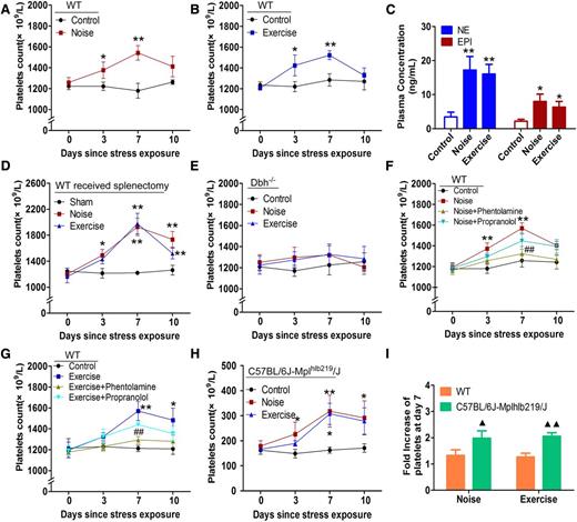
First, a gradual and significant increase in the platelet counts was observed in the peripheral blood of C57BL/6 mice suffering from either continual noise or exhaustive exercise stress for 7 days (Figure 1A-B). Interestingly, the platelet counts accumulated when mice were subjected to stress for an additional 7 days (supplemental Figure 1, available on the Blood Web site). A significant elevation in plasma NE and EPI was detected in these mice (Figure 1C). To clarify the cause of the increase in platelet counts, an additional experiment was performed using mice that had received a splenectomy. A significant increase in the platelet counts persisted in these mice after continual stresses (Figure 1D), indicating that the increase in the platelet count was probably the result of enhanced platelet production and not simply a transient release of platelets from the spleen. In contrast, the same stresses did not induce significant increases in the platelet counts in the dopamine β-hydroxylase–deficient (Dbh−/−) mice, which were defective in NE and EPI (Figure 1E). In addition, the stress-induced increase in platelet counts in WT mice could be largely blocked by an α-adrenoceptor antagonist (phentolamine) but not a β-adrenoceptor antagonist (propranolol) (Figure 1F-G). To further document the effect of stress-induced sympathetic stimulation on platelet counts, we performed the same experiments on C57BL/6J-Mplhlb219/J mice that are defective in TPO-dependent thrombopoiesis due to a homozygous mutation of the receptor of TPO, c-Mpl. We found a more apparent increase in platelet counts in these mice after stress (Figure 1H-I), indicating that sympathetic stress on thrombopoiesis is not a result of merely working through the TPO signaling axis. These results suggest that the elevated release of NE and EPI after continual stress probably contributes to the increase in platelet counts by stimulating thrombopoiesis.
Continual stress exposure elevates the peripheral platelet level. (A-B) Peripheral platelet counts in WT C57BL/6 mice subjected to noise stress or exhaustive exercise stress for 7 days. (C) High-pressure liquid chromatography (HPLC) analysis of plasma concentrations of NE and EPI in WT mice after stress. (D) Peripheral platelet counts in splenectomized mice exposed to continual noise or exhaustive exercise stress. (E) Peripheral platelet counts in Dbh−/− mice after exposure to continual noise or exhaustive exercise stress. (F-G) Peripheral platelet levels in WT mice injected intraperitoneally with saline, 15 mg/kg phentolamine, or 5 mg/kg propranolol 30 minutes prior to stress exposure. (H) Peripheral platelet counts in C57BL/6J-Mplhlb219/J mice after exposure to noise or exhaustive exercise stress. (I) Fold induction of platelet levels on day 7 in C57BL/6J-Mplhlb219/J and WT mice after exposure to noise or exhaustive exercise stress. *P < .05, **P < .01, vs control; #P < .05, ##P < .01, vs stress group; ▲P < .05, ▲▲P < .01, WT vs C57BL/6J-Mplhlb219/J mice. Each group contains 6 to 7 mice.
Continual stress exposure elevates the peripheral platelet level. (A-B) Peripheral platelet counts in WT C57BL/6 mice subjected to noise stress or exhaustive exercise stress for 7 days. (C) High-pressure liquid chromatography (HPLC) analysis of plasma concentrations of NE and EPI in WT mice after stress. (D) Peripheral platelet counts in splenectomized mice exposed to continual noise or exhaustive exercise stress. (E) Peripheral platelet counts in Dbh−/− mice after exposure to continual noise or exhaustive exercise stress. (F-G) Peripheral platelet levels in WT mice injected intraperitoneally with saline, 15 mg/kg phentolamine, or 5 mg/kg propranolol 30 minutes prior to stress exposure. (H) Peripheral platelet counts in C57BL/6J-Mplhlb219/J mice after exposure to noise or exhaustive exercise stress. (I) Fold induction of platelet levels on day 7 in C57BL/6J-Mplhlb219/J and WT mice after exposure to noise or exhaustive exercise stress. *P < .05, **P < .01, vs control; #P < .05, ##P < .01, vs stress group; ▲P < .05, ▲▲P < .01, WT vs C57BL/6J-Mplhlb219/J mice. Each group contains 6 to 7 mice.
NE and EPI exert a stimulatory effect on thrombopoiesis in vivo and promote platelet recovery in mice with thrombocytopenia induced by irradiation
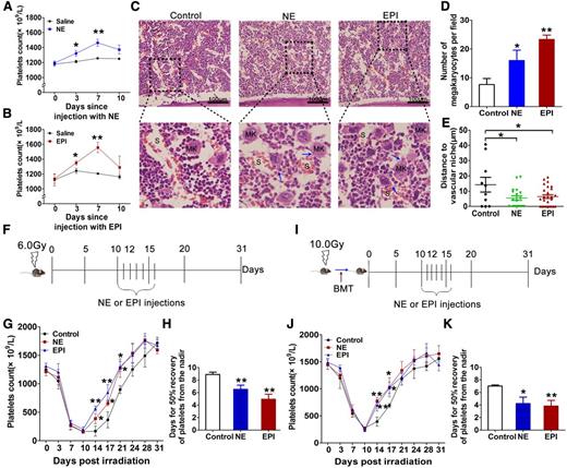
Subsequently, we treated C57BL/6 mice with NE or EPI for 7 days to verify their roles in thrombopoiesis facilitation in vivo. As expected, peripheral platelets gradually increased in the mice after NE and EPI treatments. On day 7, the platelet levels were ∼1.35-fold and 1.45-fold greater than baseline, respectively (Figure 2A-B). H&E staining revealed that the numbers of MKs in the BM in the NE- and EPI-treated mice were significantly increased (Figure 2C-D). More MKs were in close proximity to the vascular sinusoids, and some of them extended their pseudopods into the space between the endothelial cell junctions (Figure 2C). The mean distance from the MKs to the vascular sinusoids in NE- and EPI-treated mice was reduced compared with that in control mice (Figure 2E). These data demonstrate that NE and EPI stimulation can promote thrombopoiesis in vivo.
NE and EPI administration elevate platelet levels in vivo. (A-B) Changes in the peripheral platelet levels in normal C57BL/6 mice injected with saline, NE, or EPI once a day for 7 days. (C) Distributions of MKs in the BM (H&E staining) in the mice. Arrows point to the pseudopods of MKs (proplatelets) inserting into the vascular sinusoids. (D) Quantification of MKs in the BM in the mice. (E) Quantification of the distance between MKs and the vascular sinusoids in the BM. (F) Experimental schematic of NE and EPI administration in C57BL/6 mice subjected to irradiation. (G) Changes in the peripheral platelet counts in irradiated mice receiving NE or EPI treatment. (H) Time (days) needed for 50% recovery of platelet loss from the nadir in irradiated mice receiving NE or EPI treatment. (I) Experimental schematic of NE and EPI administration in C57BL/6 mice that received BMT after irradiation. (J) Changes in the peripheral platelet counts in grafted mice that received NE or EPI treatment. (K) Time (days) needed for 50% recovery of platelet loss from the nadir in grafted mice receiving NE or EPI treatment. *P < .05, **P < .01, vs control. Each group contains 6 to 7 mice.
NE and EPI administration elevate platelet levels in vivo. (A-B) Changes in the peripheral platelet levels in normal C57BL/6 mice injected with saline, NE, or EPI once a day for 7 days. (C) Distributions of MKs in the BM (H&E staining) in the mice. Arrows point to the pseudopods of MKs (proplatelets) inserting into the vascular sinusoids. (D) Quantification of MKs in the BM in the mice. (E) Quantification of the distance between MKs and the vascular sinusoids in the BM. (F) Experimental schematic of NE and EPI administration in C57BL/6 mice subjected to irradiation. (G) Changes in the peripheral platelet counts in irradiated mice receiving NE or EPI treatment. (H) Time (days) needed for 50% recovery of platelet loss from the nadir in irradiated mice receiving NE or EPI treatment. (I) Experimental schematic of NE and EPI administration in C57BL/6 mice that received BMT after irradiation. (J) Changes in the peripheral platelet counts in grafted mice that received NE or EPI treatment. (K) Time (days) needed for 50% recovery of platelet loss from the nadir in grafted mice receiving NE or EPI treatment. *P < .05, **P < .01, vs control. Each group contains 6 to 7 mice.
We then evaluated the potential effects of NE and EPI stimulation in promoting thrombopoiesis in mice with severe thrombocytopenia. C57BL/6 mice were total-body irradiated with a single dose of 6.0 Gy. Then, NE or EPI was administered for 7 days starting on the 10th day when the platelet level reached the nadir (Figure 2F). Both the NE and EPI treatments significantly increased the recovery of platelets in the irradiated mice (Figure 2G), and the time needed for 50% recovery of platelet loss from the nadir moved forward by 2.37 ± 0.94 days and 3.97 ± 0.74 days, respectively (Figure 2H). Additionally, we assessed the effects of NE and EPI treatment on platelet recovery in C57BL/6 mice that received BM transplantation (BMT) following a 10.0 Gy lethal irradiation (Figure 2I) and obtained a similar result (Figure 2J-K). These data indicate that sympathetic stimulation is favorable to platelet recovery in vivo after suffering from severe radiation.
Sympathetic stimulation-enhanced thrombopoiesis due to continual stress may contribute to the aggravation of atherosclerosis in ApoE−/− mice
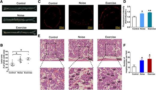
Because platelets play crucial roles not only in hemostasis but also in thrombosis under different conditions, we investigated the influence of a sympathetic stimulation-induced platelet increase on atherothrombosis. ApoE−/− mice suffering from noise or exhaustive exercise stress for 8 weeks developed more severe atherosclerotic lesions (Figure 3A). Quantitative analysis revealed that the areas of the atherosclerotic aortic plaques were much larger in the stress-stimulated mice (Figure 3B). Furthermore, a collar-induced atherosclerosis model was applied to estimate the accumulation of platelets in the atherosclerotic lesions,24 and more platelets were detected in the carotid artery in the stress-stimulated mice (Figure 3C-D). Correspondingly, the numbers of MKs in the BM were much higher in the stress-stimulated ApoE−/− mice than those in the control ApoE−/− mice (Figure 3E-F). Based on our findings, we conclude that sympathetic stress-stimulated thrombopoiesis may also contribute to the aggravation of atherothrombosis.
Long-term stress-induced thrombopoiesis contributes to the aggravation of atherosclerosis in ApoE−/− mice. (A) Representative aortas from ApoE−/− mice subjected to noise or exhaustive exercise stress for 8 weeks. (B) Quantification of the lesion size in the whole aorta. Each data point represents a value from a single mouse; n = 6. (C) Immunofluorescence staining of platelets with the CD41 antibody in the atherosclerotic lesions in the right carotid artery. (D) Quantification of fluorescence intensity of CD41 in the atherosclerotic lesions in the right carotid artery. (E) Distributions of MKs in the BM in the mice. (F) Quantification of MKs in the BM. *P < .05, **P < .01, vs control.
Long-term stress-induced thrombopoiesis contributes to the aggravation of atherosclerosis in ApoE−/− mice. (A) Representative aortas from ApoE−/− mice subjected to noise or exhaustive exercise stress for 8 weeks. (B) Quantification of the lesion size in the whole aorta. Each data point represents a value from a single mouse; n = 6. (C) Immunofluorescence staining of platelets with the CD41 antibody in the atherosclerotic lesions in the right carotid artery. (D) Quantification of fluorescence intensity of CD41 in the atherosclerotic lesions in the right carotid artery. (E) Distributions of MKs in the BM in the mice. (F) Quantification of MKs in the BM. *P < .05, **P < .01, vs control.
NE and EPI have the ability to expand CD34+ cells but cannot promote the commitment to MK lineage and proliferation of MKs
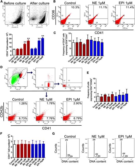
Next, we performed a series of experiments in vitro to investigate the direct effects of NE and EPI stimulation on thrombopoiesis. First, we found that either NE or EPI exposure could increase the numbers of CD34+ cells in a dose-dependent manner. (Figure 4A), which was consistent with the previous study reported by Spiegel et al.10 However, our data showed that NE and EPI had no significant effects on the commitment of CD34+ cells to megakaryocyte-erythroid progenitor lineage (Figure 4B-C) and the differentiation of MK (Figure 4D-E). Moreover, both NE and EPI were unable to increase the number and DNA content of primary MKs (Figure 4F-G). These data suggest that although NE and EPI can expand CD34+ cells, they do not promote the commitment of CD34+ cells to MK lineage and the proliferation and differentiation of MKs.
NE and EPI expand the population of CD34+ cells but have no significant effect on the commitment to MK lineage and the proliferation of MKs. Human cord blood–derived CD34+ cells were cultured with NE and EPI together with different cytokines for 7 days. (A) Flow cytometric analysis of the numbers of CD34+ cells labeled with APC-conjugated anti-CD34 antibody after the cells being treated with NE and EPI at the indicated concentrations in the presence of 20 ng/mL rhSCF. (B) The expression of CD36+CD41+ on CD34+ cells analyzed by flow cytometric analysis. (C) Mean frequency of CD34+ cells expressing CD36+CD41+ after the cells being treated with different concentrations of NE or EPI in the presence of rhSCF. (D) Flow cytometric analysis of CD41+CD42b+ expression on the cells after treatment with NE (1 μM) or EPI (1 μM) in the presence of 20 ng/mL rhSCF. (E) Mean frequency of cells expressing CD41+CD42b+ after CD34+ cells being treated with different concentrations of NE or EPI in the presence of 20 ng/mL rhSCF. (F) Flow cytometric analysis of CD41+ cell counts after NE and EPI treatment at the indicated concentrations in the presence of 20 ng/mL rhTPO. (G) Histogram of DNA content in CD41+ cells after CD34+ cells being cultured with rhTPO and double-labeled with FITC-conjugated CD41 and PI. *P < .05, **P < .01 vs control. SCF, stem cell factor.
NE and EPI expand the population of CD34+ cells but have no significant effect on the commitment to MK lineage and the proliferation of MKs. Human cord blood–derived CD34+ cells were cultured with NE and EPI together with different cytokines for 7 days. (A) Flow cytometric analysis of the numbers of CD34+ cells labeled with APC-conjugated anti-CD34 antibody after the cells being treated with NE and EPI at the indicated concentrations in the presence of 20 ng/mL rhSCF. (B) The expression of CD36+CD41+ on CD34+ cells analyzed by flow cytometric analysis. (C) Mean frequency of CD34+ cells expressing CD36+CD41+ after the cells being treated with different concentrations of NE or EPI in the presence of rhSCF. (D) Flow cytometric analysis of CD41+CD42b+ expression on the cells after treatment with NE (1 μM) or EPI (1 μM) in the presence of 20 ng/mL rhSCF. (E) Mean frequency of cells expressing CD41+CD42b+ after CD34+ cells being treated with different concentrations of NE or EPI in the presence of 20 ng/mL rhSCF. (F) Flow cytometric analysis of CD41+ cell counts after NE and EPI treatment at the indicated concentrations in the presence of 20 ng/mL rhTPO. (G) Histogram of DNA content in CD41+ cells after CD34+ cells being cultured with rhTPO and double-labeled with FITC-conjugated CD41 and PI. *P < .05, **P < .01 vs control. SCF, stem cell factor.
NE and EPI exposure significantly increase the adhesion and migration of MKs via α2-adrenoceptor-mediated ERK1/2 activation
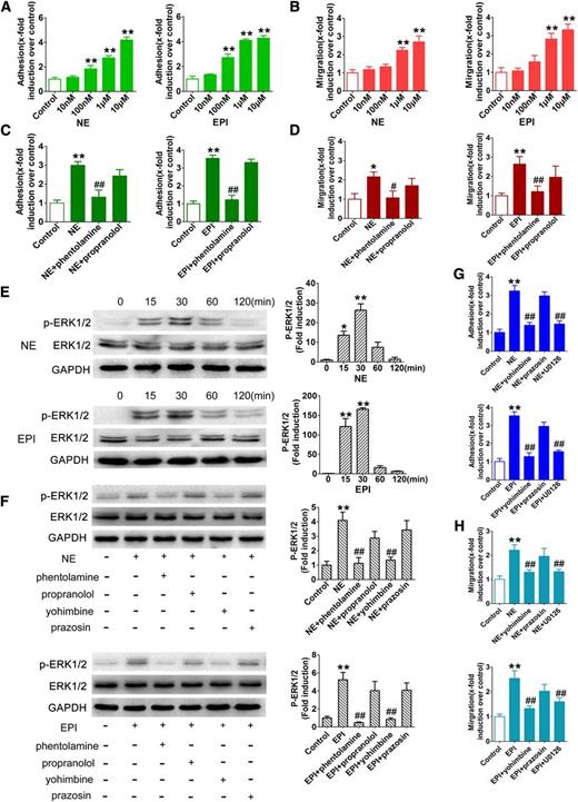
During the process of thrombopoiesis in vivo, MKs move from the osteoblastic niche to the vascular niche. The cellular behaviors of adhesion and migration are crucial for MK maturation and platelet production.6 Notably, we found that both NE and EPI promoted MK adhesion in a dose-dependent manner (Figure 5A; supplemental Figure 2). Similarly, both NE and EPI had a strong ability to promote MK migration (Figure 5B; supplemental Figure 3). The ability of NE and EPI to induce MK adhesion and migration could be largely inhibited by an α- but not a β-adrenoceptor antagonist (Figure 5C-D; supplemental Figures 2-3), although both the α- and β-adrenoceptor subtypes were expressed in MKs (supplemental Figure 4). Further investigation revealed that both NE and EPI stimulation rapidly increased the phosphorylation of ERK1/2 (Figure 5E), which could be inhibited by the α2-adrenoceptor antagonist yohimbine but not the α1-adrenoceptor antagonist prazosin (Figure 5F). Correspondingly, pretreatment with the ERK1/2-specific inhibitor U0126 significantly inhibited the adhesion and migration of MKs induced by NE and EPI in a manner that was similar to the effect of yohimbine (Figure 5G-H; supplemental Figures 2-3). These results demonstrate that NE and EPI stimulation can enhance the adhesion and migration of MKs through α2-adrenoceptor–mediated ERK1/2 activation.
NE and EPI promote the adherence and migration of MKs. The progeny of CD34+ cells cultured with rhTPO for 13 days were purified first and then used for the experiments. (A-B) Adherent and migrated MKs treated with different concentrations of NE or EPI. (C-D) Adherence and migration of MKs pretreated with phentolamine (10 μM) or propranolol (10 μM) for 20 minutes before incubation with 1 μM NE or EPI. (E) Western blot analysis for ERK1/2 phosphorylation in MKs after exposure to NE (1 μM) or EPI (1 μM) for the indicated times. (F) ERK1/2 phosphorylation in MKs pretreated with different adrenergic antagonists for 20 minutes followed by NE (1 μM) or EPI (1 μM) treatment of 30 minutes. (G-H) The inhibitory effect of yohimbine, prazosin, and U0126 on the adherence and migration of MKs induced by NE (1 μM) or EPI (1 μM). *P < .05, **P < .01, vs control; #P < .05, ##P < .01, vs NE or EPI treatment group.
NE and EPI promote the adherence and migration of MKs. The progeny of CD34+ cells cultured with rhTPO for 13 days were purified first and then used for the experiments. (A-B) Adherent and migrated MKs treated with different concentrations of NE or EPI. (C-D) Adherence and migration of MKs pretreated with phentolamine (10 μM) or propranolol (10 μM) for 20 minutes before incubation with 1 μM NE or EPI. (E) Western blot analysis for ERK1/2 phosphorylation in MKs after exposure to NE (1 μM) or EPI (1 μM) for the indicated times. (F) ERK1/2 phosphorylation in MKs pretreated with different adrenergic antagonists for 20 minutes followed by NE (1 μM) or EPI (1 μM) treatment of 30 minutes. (G-H) The inhibitory effect of yohimbine, prazosin, and U0126 on the adherence and migration of MKs induced by NE (1 μM) or EPI (1 μM). *P < .05, **P < .01, vs control; #P < .05, ##P < .01, vs NE or EPI treatment group.
NE and EPI promote PPF in an α2-adrenoceptor/ERK1/2-RhoA GTPase-dependent pathway
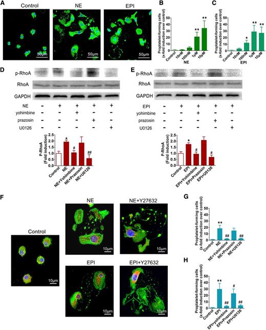
Next, we investigated the effects of NE and EPI stimulation on PPF during the late stage of thrombopoiesis. Both NE and EPI distinctly promoted PPF by mature MKs; this effect was characterized by cytoplasm extensions with cytoskeleton reorganization and the formation of pseudopods and bead-like structures (Figure 6A). Further observation showed that both NE and EPI could stimulate MKs to undergo PPF in a dose-dependent manner (Figure 6B-C). To uncover the underlying mechanisms, we investigated the influences of NE and EPI on the activity of RhoA GTPase, which was reported to regulate the rearrangement of the MK cytoskeleton during PPF.25,26 Both the NE and EPI treatments led to a significant increase in the Ser188 phosphorylation of RhoA (Figure 6D-E). Ser188 phosphorylation negatively regulates the activity of RhoA27,28 ; thus, our results indicated that NE and EPI stimulated PPF via the downregulation of RhoA activity. The role of RhoA in NE- and EPI-induced PPF was confirmed by the finding that Rho kinase (ROCK; a downstream effector of RhoA) inhibition could facilitate the elongation of the pseudopodia of MKs treated with NE and EPI (Figure 6F). Notably, pretreatment with yohimbine and U0126 almost completely inhibited the NE- and EPI-induced increase in RhoA phosphorylation (Figure 6D-E). Concordantly, U0126 significantly inhibited NE- and EPI-induced PPF in a manner similar to the effect of yohimbine (Figure 6G-H). These data suggest that NE and EPI stimulation can promote PPF via an α2-adrenoceptor/ERK1/2-RhoA GTPase-dependent pathway.
NE and EPI facilitate PPF. (A) Representative photographs of PPF after NE (1 μM) or EPI (1 μM) stimulation. Cytoskeleton actin (green) and the nucleus (blue) were stained. (B-C) Quantification of proplatelet-forming MKs treated with NE or EPI at different concentrations under an inverted microscope. (D-E) Western blot analysis of the expression of the phospho-RhoA in MKs prestimulated with yohimbine, prazosin, or U0126 for 20 minutes followed by NE or EPI treatment of 30 minutes. (F) Morphology exhibition of MKs treated with 10 μM of the ROCK inhibitor Y27632 for 20 minutes before stimulation with NE or EPI. Cytoskeleton actin (green), tubulin (red), and the nucleus (blue) were stained. (G-H) The counts of proplatelet-forming MKs exposed to 10 μM prazosin, 10 μM yohimbine, or 10 μM U0126 prior to NE or EPI stimulation. *P < .05, **P < .01, vs control; #P < .05, ##P < .01, vs NE or EPI treatment group.
NE and EPI facilitate PPF. (A) Representative photographs of PPF after NE (1 μM) or EPI (1 μM) stimulation. Cytoskeleton actin (green) and the nucleus (blue) were stained. (B-C) Quantification of proplatelet-forming MKs treated with NE or EPI at different concentrations under an inverted microscope. (D-E) Western blot analysis of the expression of the phospho-RhoA in MKs prestimulated with yohimbine, prazosin, or U0126 for 20 minutes followed by NE or EPI treatment of 30 minutes. (F) Morphology exhibition of MKs treated with 10 μM of the ROCK inhibitor Y27632 for 20 minutes before stimulation with NE or EPI. Cytoskeleton actin (green), tubulin (red), and the nucleus (blue) were stained. (G-H) The counts of proplatelet-forming MKs exposed to 10 μM prazosin, 10 μM yohimbine, or 10 μM U0126 prior to NE or EPI stimulation. *P < .05, **P < .01, vs control; #P < .05, ##P < .01, vs NE or EPI treatment group.
NE and EPI promote platelet release and activation through the ERK1/2 signal pathway
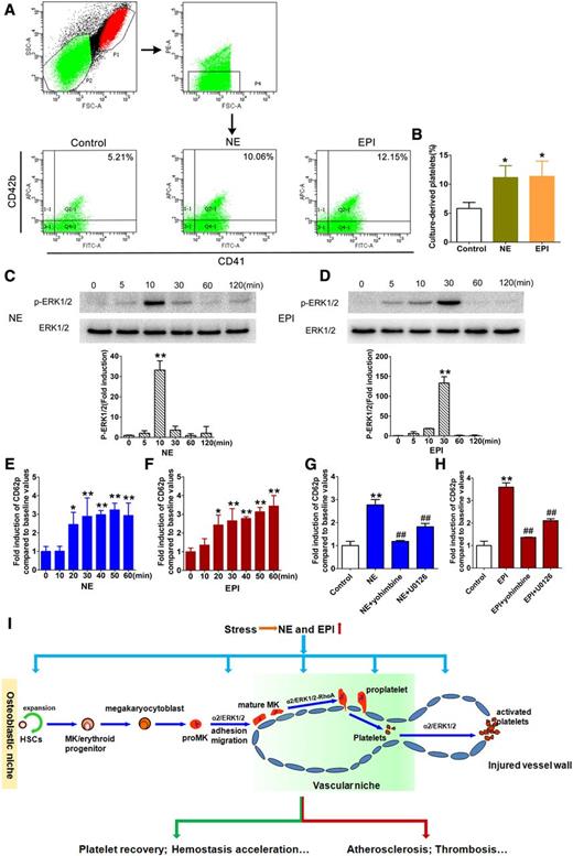
Finally, we investigated the effects of NE and EPI stimulations on platelet production by terminally differentiated MKs. As assessed by flow cytometry, both NE and EPI stimulate platelet release from MKs (Figure 7A-B). However, the increased platelets induced by NE and EPI are incapable of exacerbating atherothrombosis until they are activated. Interestingly, previous studies reported the ability of NE and EPI to trigger platelet activation after α2-adrenoceptor activation,29,30 but the underlying mechanism was unclear. Here, apart from the fact that NE and EPI markedly induced platelet activation as reflected by increased surface expression of CD62P,15 we revealed that ERK1/2 phosphorylation was preceded by the peak of platelet activation (Figure 7C-F), and ERK1/2 inhibition could significantly inhibit NE- and EPI-induced platelet activation in a manner similar to the effect of yohimbine (Figure 7G-H; supplemental Figure 5). These findings indicate that NE and EPI stimulations can also promote platelet production and activation via an ERK1/2-dependent pathway mediated by the α2-adrenoceptor.
NE and EPI promote platelet production and activation. (A) Flow cytometric analysis of platelet production from MKs induced by NE or EPI. CD34+ cells were cultured with rhTPO for 13 days and then treated with NE (1 μM) or EPI (1 μM) for 24 hours. (B) Mean percentages of culture-derived platelets induced by NE or EPI. (C-D) Western blot analysis of ERK1/2 phosphorylation in isolated platelets from the health donors treated with NE or EPI for different times. (E-F) Flow cytometry analysis of the time-course changes of in CD62p expression in isolated platelets induced by NE (10 μM) or EPI (10 μM). (G-H) CD62p expression in isolated platelets treated with yohimbine (10 μM) and U0126 (10 μM) before NE and EPI stimulation. (I) Schematic illustration of the role of sympathetic stress in regulating thrombopoiesis and its pathophysiological implications. The elevated NE and EPI levels induced by sympathetic stress stimulate thrombopoiesis and platelet activation via α2-adrenoceptor–mediated ERK1/2 signaling. As a result, the increased platelet counts and activation promote 2 different outcomes (hemostasis facilitation or thrombosis aggravation) under different pathophysiological conditions. *P < .05, **P < .01, vs control; #P < .05, ##P < .01, vs NE or EPI treatment group.
NE and EPI promote platelet production and activation. (A) Flow cytometric analysis of platelet production from MKs induced by NE or EPI. CD34+ cells were cultured with rhTPO for 13 days and then treated with NE (1 μM) or EPI (1 μM) for 24 hours. (B) Mean percentages of culture-derived platelets induced by NE or EPI. (C-D) Western blot analysis of ERK1/2 phosphorylation in isolated platelets from the health donors treated with NE or EPI for different times. (E-F) Flow cytometry analysis of the time-course changes of in CD62p expression in isolated platelets induced by NE (10 μM) or EPI (10 μM). (G-H) CD62p expression in isolated platelets treated with yohimbine (10 μM) and U0126 (10 μM) before NE and EPI stimulation. (I) Schematic illustration of the role of sympathetic stress in regulating thrombopoiesis and its pathophysiological implications. The elevated NE and EPI levels induced by sympathetic stress stimulate thrombopoiesis and platelet activation via α2-adrenoceptor–mediated ERK1/2 signaling. As a result, the increased platelet counts and activation promote 2 different outcomes (hemostasis facilitation or thrombosis aggravation) under different pathophysiological conditions. *P < .05, **P < .01, vs control; #P < .05, ##P < .01, vs NE or EPI treatment group.
Discussion
Sympathetic regulation has attracted an increasing amount of attention because it plays important roles in the body, including influencing on the fate and activity of certain hematopoietic cells.8,10,31 In this study, we show for the first time that the sympathetic transmitters NE and EPI have stimulatory effects on thrombopoiesis and demonstrate that continual and intensive stress may increase platelet production in vivo through sympathetic activation (Figure 7I). Our findings not only deepen the understanding of how sympathetic regulation affects hematopoiesis but also provide new insights into the physiopathological implications of sympathetic stress.
The process of thrombopoiesis includes several consecutive stages.4 Previous studies were primarily focused on the early and middle stages of thrombopoiesis, including the commitment to MK lineage and the MK proliferation.20,32 Here, we demonstrate that the sympathetic transmitters NE and EPI stimulate platelet production by promoting MK adhesion, migration, PPF and platelet release, in addition to the expansion of CD34+ cells. These findings are consistent with our recent study that showed that human growth hormone treatment resulted in a quick increase in platelet production by regulating the late stage of thrombopoiesis, including the promotion of PPF and platelet release. Although the increase in platelet counts induced by sympathetic stimulation is modest, we suspect there is a possibility for the combined application of sympathetic stimulation with hematopoietic growth factors to the quick recovery of platelets in patients with thrombocytopenia.
The physiological and pathological effects of sympathetic stimulation are typically dependent on the activation of adrenergic receptors.33,34 Previous studies have demonstrated that sympathetic stimulation expands the HSC population via β2-adrenergic receptor activation,10 whereas β3-adrenergic receptor activation triggers HSC/progenitor mobilization by decreasing stromal cell-derived factor-1 (or CXC chemokine ligand 12) messenger RNA levels in BM stromal cells.31 Activation of the β2-adrenergic receptor is involved in the isoproterenol-induced proliferation of erythroid progenitor cells,35 whereas α1-adrenergic receptor activation mediates the stimulatory effect of noradrenergic regulation on lymphohematopoiesis.12 In the present study, we found that blockade of the α2-adrenergic receptor could largely inhibit the actions of NE and EPI in promoting MK adhesion, migration, PPF, platelet release, and platelet activation, indicating that the stimulatory effect of NE and EPI on thrombopoiesis is mediated by the activation of the α2-adrenergic receptor. These findings demonstrate that sympathetic innervation is involved in various aspects of hematopoiesis via activation of different adrenergic receptors, and the activation of the α2-adrenergic receptor is preferred for thrombopoiesis.
Then, we revealed that ERK1/2 is the key mediator downstream of the α2-adrenergic receptor signaling pathway in NE and EPI-induced MK adhesion and migration, which was consistent with previous reports that ERK1/2 signaling is involved in the adherence and mobility of various cell types.20,32,36 Furthermore, ERK1/2 activation downregulates the RhoA activity in mature MKs upon treatment with NE or EPI. These effects led to the rearrangement of the actin and microtublin cytoskeletons, which was a known key determinant for PPF, platelet formation, and platelet release.25,37 Although both NE and EPI stimulate platelet activation, the underlying mechanism is unclear. Here, we discovered that NE- and EPI-induced platelet activation was at least partially attributed to ERK1/2 signaling. Therefore, we conclude that ERK1/2 signaling following α2-adrenergic receptor activation plays a crucial role in NE- and EPI-stimulated thrombopoiesis and platelet activation.
It has been recognized for many years that peripheral platelet counts may increase when the body is subjected to intensive external or internal stresses.38,39 However, the underlying mechanisms have not been fully elucidated. In our study, we found there was still an evident elevation of platelet levels in splenectomized mice after long-term stresses. Therefore, we propose that the increase in platelet counts is primarily due to the enhanced platelet production by MKs, which is supported by the stimulatory effect of NE and EPI on thrombopoiesis that is observed both in vitro and in vivo. However, several previous reports noted that the increase in circulating platelets shortly after each single exercise might be attributed to the rapid release of platelets from the spleen.40,41 The discrepancy may be due to the different type and duration of the stress performed in different studies. Normally, the course of thrombopoiesis requires several days, and short-term stimulations should not induce significant platelet production. Indeed, we observed an even higher and longer elevation of platelet counts in mice that had undergone a splenectomy after long-term stress treatment, which may be explained by the deficiency in the splenic elimination of platelets.42 Additionally, adrenergic stresses were found to induce a more apparent increase in the platelet counts in mice with the c-Mpl homozygous mutation, probably because TPO/c-Mpl does not work on terminal MK maturation and even negatively regulate platelet shedding through Janus kinase–signal transducer and activator of transcription signaling activation.43-45 This finding also suggests that the sympathetic stimulation may play an important role in maintaining thrombopoiesis when the TPO/c-Mpl–associated pathway is defective.
Because platelets play important roles in both hemostasis and thrombosis, there may be a dual outcome of the enhanced thrombopoiesis facilitated by sympathetic stimulations (Figure 7I). Previous studies have shown that adrenergic stresses are harmful to the cardiovascular system because they hyperstimulate the activity of platelets.46,47 In this study, we found that long-term stresses aggravated atherothrombosis in ApoE−/− mice, accompanied by increased production of platelets by MKs. Although we cannot measure the relative contribution of the increase in the platelet count and platelet activation to atherothrombosis, it is undeniable that the increased number of platelets caused by adrenergic stress may increase the risk of atherothrombosis because the platelet count is also closely related to cardiovascular disease.48,49 Thus, our study also provides new insights into the pathogenesis of thrombosis induced by continual physical and/or psychological stress.
The online version of this article contains a data supplement.
The publication costs of this article were defrayed in part by page charge payment. Therefore, and solely to indicate this fact, this article is hereby marked “advertisement” in accordance with 18 USC section 1734.
Acknowledgments
The authors thank Michelle Meredyth-Stewart for grammar revision of this manuscript and helpful discussions, Xiaolan Fu for flow cytometric analysis assistance, and Xiangyu Ma and Yao Zhang for data analysis.
This work was supported by grants from the National Natural Science Fund of China (no. 81573084, 31500950), the Fund from People’s Liberation Army (BSW11J009), and the Funds of Key Laboratory of Trauma, Burn and Combined Injury (no. SKLZZ201115, SKLZZ201016, and SKLKF200911).
Authorship
Contribution: S.C. performed experiments, analyzed data, and wrote the paper; C.D. performed experiments, analyzed data, and participated in manuscript preparation; G.Z., M.S., and Y.X. contributed to animal experiments and data analysis; K.Y., X.W., D.Z., and F.C. contributed to the in vitro experiments and image scoring; S.W., M.C., C.W., and T.H. contributed to the in vivo experiments and performed a portion of the in vitro experiments; F.W., A.W., and F.L. contributed to the initial experimental design; T.C. and Y.S. discussed and edited the manuscript; and J.Z. and J.W. conceived and supervised the study, analyzed the data, and wrote and revised the manuscript.
Conflict-of-interest disclosure: The authors declare no competing financial interests.
Correspondence: Junping Wang, State Key Laboratory of Trauma, Burns and Combined Injury, Institute of Combined Injury, College of Preventive Medicine, Third Military Medical University, Gaotanyan St, 30 Chongqing 400038, China; e-mail: wangjunping@tmmu.edu.cn or wangjunp@yahoo.com; and Jinghong Zhao, Department of Nephrology, Xinqiao Hospital, Third Military Medical University, Xinqiao St, 183 Chongqing, 400037, China; e-mail: zhaojh@tmmu.edu.cn.
References
Author notes
S.C. and C.D. contributed equally to this work.

