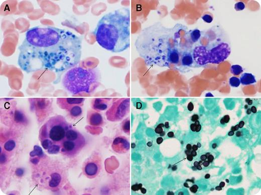
A 21-year-old man with Crohn disease and taking multiple immunosuppressive drugs presented with fevers, chills, night sweats, epistaxis, and dyspnea. He had bicytopenia (hemoglobin 9.3 g/dL, platelets 12 × 109/L, and normal leukocyte count). Biochemical studies showed markedly elevated ferritin (15 140 µg/L), decreased fibrinogen (159 mg/dL), and elevated D-dimer (2735 ng/mL) and aspartate aminotransferase (76 U/L). Computed tomography showed bilateral pulmonary micronodular infiltrates and splenomegaly. Bone marrow (BM) aspirate (Wright-Giemsa stain; panels A-B) and biopsy (hematoxylin and eosin stain; panel C) demonstrated hemophagocytosis with numerous histiocytes containing yeast forms (arrows) and hematopoietic precursors. Grocott’s methenamine silver stain highlights the yeasts (panel D). Blood, BM, and bronchoalveolar lavage cultures confirmed Histoplasma capsulatum. The patient was treated with antifungal therapy with subsequent recovery.
Hemophagocytic lymphohistiocytosis (HLH) is a systemic syndrome of histiocytic activation with hypercytokinemia. Acquired HLH is commonly associated with infections, malignancies, and collagen vascular disorders. HLH developed in this patient secondary to disseminated histoplasmosis in the setting of iatrogenic immunosuppression. HLH is associated with high mortality without appropriate treatment, and early intervention is imperative. BM examination, in conjunction with clinical and laboratory findings, is crucial for timely intervention. A diligent BM search for fungal microorganisms is also warranted in immunocompromised HLH patients because BM can sometimes be the only location to obtain the diagnosis.
For additional images, visit the ASH IMAGE BANK, a reference and teaching tool that is continually updated with new atlas and case study images. For more information visit http://imagebank.hematology.org.


This feature is available to Subscribers Only
Sign In or Create an Account Close Modal