Abstract
Tumor suppressor P53 regulates multiple signaling pathways triggered by diverse cellular stresses including DNA damages, oncogenic stimulations, and hypoxic stress, resulting in cell-cycle arrest, apoptosis, and senescence. P53 signaling is also important for double-stranded DNA breaks (DSBs) induced during physiologic events, i.e., rearrangement of antigen-specific receptors. It has been reported that P53-mediated DSB checkpoint contribute to normal murine T lymphopoiesis, especially at the double-negative (DN) stage which is defined as CD4-CD8- fraction in thymus and requires rearrangements of the T cell receptor (TCR) b locus and successful pre-TCR signaling (Guidos CJ et al., Genes Dev, 1996; Jiang D et al., J Exp Med 2006; HaksMC et al., Immunity, 1999). Here we defined the role of P53 on hematopoietic development, especially lymphopoiesis, from human embryonic stem cells (ESCs).
Firstly we modified P53 gene of human ESC H1 by utilizing genome editing tool of zinc finger nuclease (ZFN) targeting the 5th exon of the P53 gene, kindly provided by Sangamo BioSciences. Sequencing analysis of the P53 knockout (KO) ES cells showed the successful deletion at the 5th exon which induced the frame shift of the downstream sequence in both of its alleles. qRT-PCR showed no stable expression of full length P53 mRNA and western blot analysis of P53 phosphorylation status in P53 KO ESCs showed undetectable levels of phosphorylated or non-phosphorylated P53 proteins when cultured in the presence or absence of apoptotic signal triggered by mitomycin C (MMC). In consistent with this, P53 KO ESCs showed significant resistance to MMC-induced cell death. In addition, P53 KO ESCs lacked apoptotic stimulation-induced upregulation of P53 downstream target genes including P53 up-regulated modulator of apoptosis (PUMA). On the other hand induction of P53 target gene P21 was not observed both in H1 and P53 KO ESCs, as reported previously by other groups (Ginis I et al., Dev Biol, 2004; Barta T et al., Stem Cells, 2010; Garc'a CP et al., Stem Cell Res, 2014; World J et al., Stem Cells, 2014).
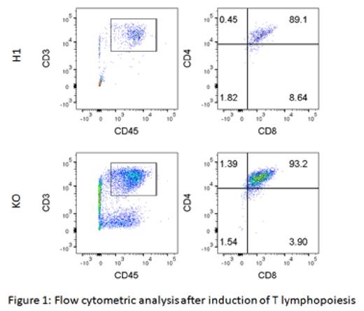
We then induced hematopoietic differentiation of P53 KO ESCs through embryoid body formation. Erythroid lineage cells developed from human ESCs were significantly suppressed in the absence of P53 signaling during embryoid body maturation. Pharmacological inhibition of P53 had the same effect as genetic disruption of P53 gene. CD34+ hematopoietic precursors were isolated from embryoid bodies originated from H1 and P53 KO ECSs, plated on OP9-DL1 stromal cells, and cultured in the presence of stem cell factor (SCF), FLT3 ligand, and interleukin (IL)-7. After 3-4 weeks of culture, CD45+CD3+ T lineage cells were induced from both H1 and P53 KO ECSs-derived CD34+ cells. Among these cells, most of the cells were in CD4+CD8+ double-positive (DP) stage, with increase in the yield of DP cells in the absence of P53 signaling (H1: 343 cells/1 x 106 input CD34+ cells; P53 KO: 2476 cells/1 x 106 input CD34+ cells; Figure). Whether pharmacological inhibition of P53 had the similar effect on T lymphopoiesis as genetic disruption of P53 gene needs to be investigated furthermore.
Our data indicate that P53 mediated signaling regulate in vitro early T lymphopoiesis from human pluripotent stem cells, especially at the transition from double negative into DP stage. These observations promoted us to perform high throughput transcriptome analysis including cDNA microarray analysis between early T lineage cells derived from H1 and P53 KO ESCs. Genes associated with the early T lymphopoiesis from human ESCs were identified and currently under further characterization.
No relevant conflicts of interest to declare.
Author notes
Asterisk with author names denotes non-ASH members.


This feature is available to Subscribers Only
Sign In or Create an Account Close Modal