Abstract
In recent years, evidence has been provided that natural killer (NK) cells can function as anti-leukemic effector cells. In humans, two NK cell populations are usually distinguished: CD56dimCD16+ NK cells form the predominant population in blood while the CD56brightCD16- NK cell population is more prominent in tissues. However, little data regarding tissue specific characteristics of human NK cells are available, especially in bone marrow as an important localization of leukemic cells. Therefore, we evaluated the expression of chemokine receptors and adhesion molecules on NK cells in healthy donor blood, bone marrow and spleen by flow cytometry.
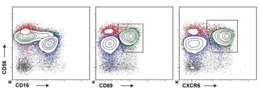
Besides the two conventional NK cell subsets, a major third NK cell population was identified in bone marrow and spleen based on the combined expression of CD69 and CXCR6 (Figure 1). CD69+CXCR6+ NK cells represented 9-50% (mean 30%) of NK cells in marrow (n=15) and 26-57% (mean 43%) of NK cells in spleen (n=7). This CD69+CXCR6+ population was not detected in blood (n=15) nor in cord blood, neither mobilized into the blood stream after G-CSF treatment. The ratio and phenotype of the remaining conventional CD56bright and CD56dim NK cells in bone marrow and spleen were comparable to blood. Early after pediatric hematopoietic stem cell transplantation, CD69+CXCR6+ NK cells were absent in bone marrow, but gradually reached normal levels within the first year after transplantation.
CD56 was expressed on marrow and spleen CD69+CXCR6+ NK cells at slightly lower levels than the conventional CD56bright NK cells and CD16 was expressed by 7-30% of CD69+CXCR6+ NK cells. CD69+CXCR6+ NK cells expressed high levels of the adhesion molecule CD54 (ICAM-1) as well as natural cytotoxicity triggering receptor NKp46 compared to the conventional CD56bright and CD56dim NK cell populations. CD69+CXCR6+ NK cells did not express the early differentiation markers CD117 (c-kit) and CD127 (IL7Rα), which are expressed by immature NK cells, type III innate lymphoid cells and, to some extent, by conventional CD56bright NK cells. The inhibitory receptor NKG2A, which is acquired early in maturation but lost during the differentiation of CD56dim NK cells, was expressed by 60% of CD69+CXCR6+ NK cells. Furthermore, CD69+CXCR6+ NK cells did not express markers acquired late in differentiation (KIRs, CD57, KLRG1, NKG2C).
In functional experiments assessing cytokine producing capacity, CD69+CXCR6+ NK cells were comparable to the CD56dim subset, requiring the combined stimulation with IL12+IL15+IL18 to produce IFN-γ. However, with respect to cytotoxic potential, CD69+CXCR6+ NK cells more resembled the CD56bright NK cell subset; in resting state, CD69+CXCR6+ NK cells expressed perforin but not granzyme B, which was upregulated during overnight IL12+IL15 stimulation. Upon co-culture with K562 tumor cells, CD69+CXCR6+ NK cells degranulated (CD107a) at levels comparable to the conventional CD56bright and CD56dim NK cells.
In summary, we identified a distinct NK cell population in human bone marrow and spleen. These cells were a) absent in blood, b) expressed tissue retention marker CD69, and c) were present alongside the conventional NK cell subsets in marrow and spleen. Together, these findings indicate that NK cells with the discriminative CD69+CXCR6+ phenotype constitute a tissue resident NK cell subset. Furthermore, CD69+CXCR6+ NK cells did not express CD49a+, distinguishing them from the recently described liver resident NK cells (Marquardt et al, 2015). Based on their surface receptor expression profile and functional characteristics, CD69+CXCR6+ NK cells have a mature signature that differs from both conventional NK cell subsets. Additional studies in healthy individuals and patients with immuno-hematological diseases are needed to investigate the immunological function of CD69+CXCR6+ NK cells. For example, these cells could have a local immunoregulatory or effector function. Alternatively, bone marrow and spleen may form a reservoir for effector cells waiting to be released into the blood stream.
No relevant conflicts of interest to declare.
Author notes
Asterisk with author names denotes non-ASH members.


This feature is available to Subscribers Only
Sign In or Create an Account Close Modal