Key Points
Plk1 ablation activates an endomitotic checkpoint in megakaryocytes.
Plk1 deficiency in megakaryocytes results in thrombocytopenia.
Abstract
Polyploidization in megakaryocytes is achieved by endomitosis, a specialized cell cycle in which DNA replication is followed by aberrant mitosis. Typical mitotic regulators such as Aurora kinases or Cdk1 are dispensable for megakaryocyte maturation, and inhibition of mitotic kinases may in fact promote megakaryocyte maturation. However, we show here that Polo-like kinase 1 (Plk1) is required for endomitosis, and ablation of the Plk1 gene in megakaryocytes results in defective polyploidization accompanied by mitotic arrest and cell death. Lack of Plk1 results in defective centrosome maturation and aberrant spindle pole formation, thus impairing the formation of multiple poles typically found in megakaryocytes. In these conditions, megakaryocytes arrest for a long time in mitosis and frequently die. Mitotic arrest in wild-type megakaryocytes treated with Plk1 inhibitors or Plk1-null cells is triggered by the spindle assembly checkpoint (SAC), and can be rescued in the presence of SAC inhibitors. These data suggest that, despite the dispensability of proper chromosome segregation in megakaryocytes, an endomitotic SAC is activated in these cells upon Plk1 inhibition. SAC activation results in defective maturation of megakaryocytes and cell death, thus raising a note of caution in the use of Plk1 inhibitors in therapeutic strategies based on polyploidization regulators.
Introduction
Megakaryocytes are bone marrow cells specialized in the generation of platelets.1 Once committed to differentiation, megakaryocyte progenitor cells undergo several rounds of endomitotic cell cycles in which the latest stages of mitosis are aborted to render polyploid cells. After genome duplication, megakaryocytes enter mitosis by breaking down the nuclear envelope and condensing chromosomes, but they exit mitosis in the absence of complete karyokinesis generating polylobulated nuclei.1,2 This process is thought to be mediated by impaired furrow formation resulting from impaired RhoA activity and defective formation of the actomyosin ring required for cytokinesis.3,4 The increase in ploidy is a necessary component of megakaryocyte differentiation, and only high-ploidy cells are able to shed platelets efficiently. Although the reason for this requirement is not completely clear, the increase in ploidy accompanies the increase in cellular volume, which may provide cells with sufficient cytoplasmic volume to generate a large number of platelets.1,5,6
Because mitosis is aborted in its later stages, the requirement for mitotic regulators during megakaryocyte polyploidization has been a matter of debate. Earlier work in cell lines suggested defective activity of mitotic cyclins and its catalytic partner cyclin-dependent kinase 1 (Cdk1; reviewed in Zimmet and Ravid5 ). Recent expression data indicate that the mitotic machinery is normally expressed in megakaryocytes, whereas it is downregulated in endocycling cells (ie, cells that polyploidize through successive cycles in the complete absence of mitotic features such as trophoblast giant cells).7 Yet, although specific mitotic kinases such as Aurora A or Aurora B or its partners are expressed in megakaryocytes, they are dispensable for polyploidization or maturation of these cells.8-13
Polo-like kinase 1 (Plk1) is a member of the Plk family involved in centrosome maturation, formation of the mitotic spindle, microtubule-chromosome attachment, and cytokinesis.14-17 Similar to Aurora A inhibition, inhibition of Plk1 results in defective centrosome maturation and spindle formation. Because of its critical functions in the mitotic cell cycle and its expression in human tumors, Plk1 has been proposed as a cancer target, and recent clinical trials with Plk1 inhibitors have indeed shown significant therapeutic effect in hematopoietic tumors.15,18-21 These findings have been used to propose new therapeutic strategies based on the idea that inhibition of mitotic kinases, including members of the Cdk, Aurora, or Plk family, should impair growth of leukemia (eg, acute megakaryocytic leukemia cells) without affecting polyploidization or function of normal megakaryocytes,9,22 a feature that could protect patients from thrombocytopenia, a major side effect of antiproliferative therapies.
To understand the relevance of Plk1 during megakaryocyte differentiation, we have analyzed the effect of the specific genetic ablation of the murine Plk1 locus using a new conditional knockout model recently generated in our laboratory. Contrary to the inhibition of Aurora kinases, elimination of Plk1 results in strong defects in megakaryocyte maturation in vivo. Plk1 deficiency prevents megakaryocyte polyploidization as a result of the activation of the spindle assembly checkpoint (SAC) in these cells. This results in prolonged mitotic arrest and death of Plk1-null megakaryocytes, resulting in severe thrombocytopenia in vivo. These data highlight the functional relevance of the SAC in endomitotic megakaryocytes and add a note of caution in the general use of mitotic kinase inhibitors in therapeutic strategies based on polyploidization regulators.
Materials and methods
Mouse models and histopathological analysis
A 0.8-kb fragment containing the murine Plk1 exon 2 was flanked with loxP sites and followed by a PGK-neomycin phosphotransferase (neo) cassette, flanked by frt sites, for positive selection of embryonic stem cell clones. Recombinant clones were selected by southern blot and the neo cassette was removed using Flp recombinase to generate the Plk1 conditional allele [Plk1(lox)] (P.W. and G.d.C., submitted for publication). Pf4-Cre mice and the megakaryocyte-specific Cdc20 conditional knockout were described previously.12,23 All animals were maintained in a mixed 129/Sv (25%) × CD1 (25%) × C57BL/6J (50%) background. Mice were housed in the pathogen-free animal facility of the Centro Nacional de Investigaciones Oncológicas (Madrid, Spain) following the animal care standards of the institution. All animal protocols were approved by the local committee for animal care and research. Thrombopoietin (TPO 1.2 μg/mouse) was injected subcutaneously into 8- to 12-week-old mice and platelet counts were analyzed 4 days later. The following antibodies were used for immunohistochemistry in tissue sections: von Willebrand factor (VWF) (FVIII, Dako; 1:2000), phospho-histone H3 (Ser10; Millipore; 1:2000) or Plk1 (new monoclonal antibody generated in our laboratory; unpublished data). For quantification, paraffin sections stained with anti-VWF were examined using an Olympus BX51 microscope equipped with objective lenses (40/0.75, 20/0.4, 10/0.25, and 4/0.1). The relative number of pixels positive for VWF was used as a score.
Flow cytometry
Flow cytometric analysis of DNA content and surface markers of megakaryocytes were determined by staining either whole bone marrow cells or hematopoietic progenitor cultures from 0 to 5 days in presence of TPO with fluorescein isothiocyanate–conjugated anti-CD41 antibody (BD Biosciences) followed by fixation with 4% paraformaldehyde at room temperature for 30 minutes followed by staining with 2 μg/mL 4,6 diaminophenylindole (DAPI, Sigma-Aldrich).
Immunofluorescence
Lin‒ bone marrow cells were grown in Dulbecco’s modified Eagle medium (Gibco) supplemented with antibiotics and 10% fetal bovine serum and stimulated with 50 to 100 ng/mL murine TPO (PeproTech) until cells were harvested for analysis (3 days unless indicated otherwise). For immunofluorescence, cells were fixed with 4% paraformaldehyde in phosphate-buffered saline for 10 minutes at 37°C, permeabilized with phosphate-buffered saline-Triton 0.15% for 2 minutes at 37°C, blocked with 3% bovine serum albumin, and subsequently incubated during 1 to 3 hours with the primary antibody against Plk1 (unpublished data), pericentrin (Abcam 4448), Bub1 (a gift from Stephen Taylor), Mad2 (a gift from Katja Wassmann), α-tubulin (Sigma T9026), phosphorylated histone H3 (pH3; Millipore 06-570). Anticentromere antibodies (ACAs; Antibodies Incorporated 15-235) were used to stain centromeres and fluorescent-conjugated phalloidin (Sigma) was used to stain actin fibers. Matching secondary antibodies with different Alexa dyes (488, 555, 594, 647; Molecular Probes) and DAPI (Prolong Gold antifade; Invitrogen) were used for nuclei visualization. Image acquisition was performed using either a confocal ultraspectral microscope (Leica TCS-SP5) or a Leica DMI fluorescence 6000B microscope. In these assays, Taxol was used at 300 nM, the Plk1 inhibitor BI2536 at 100 nM, and reversine was used at 1 μM for inhibition of the SAC kinase Mps1.24
Time-lapse microscopy
Cells were transduced with lentiviruses or retroviruses encoding histone H2B-green fluorescent protein (GFP), lamin A-CFP (a gift from V. Andrés, CNIC, Madrid) and geminin-mCherry as reported previously.12 Transduced cells were then plated on 8-well glass-bottom dishes (Ibidi) embedded in methylcellulose (MethoCult M3231; Stem Cell Technologies) mixed 1:1 with Iscove modified Dulbecco medium containing 4% fetal bovine serum and 100 ng/mL murine TPO to minimize cell movement. Time-lapse acquisition was performed with a Leica DMI 6000B microscope equipped with a ×63/1.5 N.A. objective lens or DeltaVision RT imaging system (Applied Precision; IX70/71; Olympus) equipped with a Plan Apochromatic ×40/1.42 N.A. objective lens, and maintained at 37°C in a humidified CO2 chamber. Images were acquired every 7 minutes for up to 10 days. Quantitative analysis was performed by ImageJ software.
Transmission electron microscopy
Cells were fixed for 3 hours at room temperature in 3% glutaraldehyde (vol/vol) in 0.4 M N-2-hydroxyethylpiperazine-N′-2-ethanesulfonic acid buffer pH 7.4, washed and fixed again in aqueous 1% (wt/vol) osmium tetroxide, and embedded in Epon. Electron microscopy was performed with a JEOL 1230 transmission electron microscope, at 80 kV, on ultrathin sections of 60 nm.
Statistical analysis
Statistical analysis was carried out using Prism 5 (GraphPad). All statistical tests were performed using 2-sided, unpaired Student t tests. Data with P < .05 were considered statistically significant. In most figures, *P < .05; **P < .01; ***P < .001.
Results
Lack of Plk1 results in impaired megakaryocyte maturation
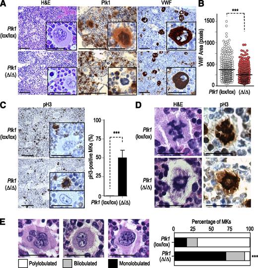
To specifically eliminate Plk1 in megakaryocytes, we made use of a Plk1 conditional allele [Plk1(lox)], recently generated in our laboratory (see “Materials and methods” for more information), and Pf4-Cre transgenic mice in which the Cre recombinase is expressed under platelet factor 4 regulatory transcriptional sequences leading to efficient recombination in medium- and high-ploidy megakaryocytes.12,23 Cre activity in Plk1(lox/lox) mice results in genetic ablation of Plk1 exon 2 generating Plk1(Δ/Δ) mice with specific ablation of Plk1 in megakaryocytes. Plk1 was absent in mitotic megakaryocytes from Plk1(Δ/Δ) mice, but was strongly expressed in control littermates (Figure 1A) or in Cdc20-deficient megakaryocytes,12 which are arrested in mitosis as a consequence of inactivation of the anaphase-promoting complex/cyclosome (APC/C; see supplemental Figure 1, available on the Blood Web site). Histopathological analysis of bone marrow sections from 8-week-old Plk1(Δ/Δ) mice indicated the presence of smaller megakaryocytes in which the amount of VWF, a marker of megakaryocyte differentiation, was significantly reduced (Figure 1A-B). In addition, these mutant bone marrows (Figure 1C), as well as Plk1(Δ/Δ) spleens (supplemental Figure 2), were characterized by the presence of abundant mitotically arrested megakaryocytes, as detected after staining with antibodies against pH3. Mitotic megakaryocytes in Plk1-deficient mice frequently presented condensed chromosomes (pH3-positive) forming typical ring structures (Figure 1D). Careful analysis of these sections revealed that megakaryocyte nuclei frequently displayed lower complexity compared with control megakaryocytes from control littermates, with a high percentage of mononucleated or binucleated megakaryocytes compared with complex polylobulated megakaryocytes typical of control bone marrows (Figure 1A,C,E).
Defective megakaryocyte maturation in the absence of Plk1. (A) Bone marrow sections from 8-week-old mice were stained with hematoxylin and eosin (H&E) or antibodies against Plk1 or the VWF. Scale bars, 100 μm (insets, 20 μm). (B) The quantification of VWF area per cell is shown in the histogram. Horizontal bars indicate the average from >500 cells per condition from at least 3 different animals. ***P < .001; Student t test. (C) Phosphorylation of pH3 in Plk1-deficient or control megakaryocytes. Scale bars, 100 μm (insets, 20 μm). The quantification of pH3-positive cells is shown in the right histogram. Data are mean ± standard deviation (SD; n = 3 mice per genotype). ***P < .001; Student t test. (D) Representative images of mitotic cells in Plk1-deficient or control mice after staining with H&E or immunodetection of pH3. Scale bars, 20 μm. (E) Analysis of nuclear complexity in Plk1-null and control megakaryocytes (MKs). Scale bars, 20 μm. In these analyses, at least 250 cells from ≥3 different animals per genotype were used. Student t test; ***P < .001.
Defective megakaryocyte maturation in the absence of Plk1. (A) Bone marrow sections from 8-week-old mice were stained with hematoxylin and eosin (H&E) or antibodies against Plk1 or the VWF. Scale bars, 100 μm (insets, 20 μm). (B) The quantification of VWF area per cell is shown in the histogram. Horizontal bars indicate the average from >500 cells per condition from at least 3 different animals. ***P < .001; Student t test. (C) Phosphorylation of pH3 in Plk1-deficient or control megakaryocytes. Scale bars, 100 μm (insets, 20 μm). The quantification of pH3-positive cells is shown in the right histogram. Data are mean ± standard deviation (SD; n = 3 mice per genotype). ***P < .001; Student t test. (D) Representative images of mitotic cells in Plk1-deficient or control mice after staining with H&E or immunodetection of pH3. Scale bars, 20 μm. (E) Analysis of nuclear complexity in Plk1-null and control megakaryocytes (MKs). Scale bars, 20 μm. In these analyses, at least 250 cells from ≥3 different animals per genotype were used. Student t test; ***P < .001.
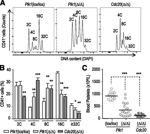
To check the effect of Plk1 ablation in megakaryocyte ploidy, bone marrow cells were isolated and their DNA content was analyzed by flow cytometry. As shown in Figure 2A, lack of Plk1 resulted in defective ploidy profiles in these CD41+ cells. Whereas control cells frequently reach 16C or 32C ploidy levels, Plk1(Δ/Δ) megakaryocytes displayed a reduced ratio of these peaks when compared with low-ploidy (4C to 8C) megakaryocytes (Figure 2B). As a control, we also analyzed megakaryocytes deficient in Cdc20, a model with reduced ploidy and severe thrombocytopenia.12 It is important to highlight that, because Plk1- and Cdc20-deficient megakaryocytes frequently accumulated as mitotic cells, these peaks at least partially represent mitotic cells (ie, 16C peaks are likely a combination of interphasic 16C cells plus mitotic 8C cells; Figure 2A-B). Plk1 deficiency resulted in increased levels of megakaryocyte progenitors (CD41+CD42‒ cells), as reported for Cdc20-deficient mice,12 but a reduced ratio of CD41+CD42+ double-positive cells, suggesting defective maturation (supplemental Figure 3). In agreement with these observations, Plk1(Δ/Δ) mice displayed a significant decrease of circulating platelets in peripheral blood (Figure 2C), suggesting that Plk1 ablation resulted in significant levels of thrombocytopenia in these mice. Platelet morphology or size was not altered in the absence of Plk1 (supplemental Figure 4). Plk1(Δ/Δ) mice, however, were resistant to treatment with TPO, suggesting a defect in the process that leads to the generation of platelets from megakaryocyte progenitors (supplemental Figure 4).
Defective megakaryocyte ploidy and circulating platelets in Plk1(Δ/Δ) mice. (A) Analysis of DNA content in gated CD41+ cells from the bone marrow of 8-week-old Plk1(Δ/Δ) or control mice. The plot is representative of 3 different animals analyzed per genotype. (B) DNA content in CD41+ cells from the bone marrow of mice with the indicated genotypes. Data are mean ± SD (n = 4 mice per genotype). *P < 0.05; **P < .01; ***P < .001; Student t test. (C) Number of circulating platelets in Plk1(Δ/Δ) or control mice. As a comparison, the average count is ∼100 × 109 platelets/L in Cdc20-deficient mice.12 ***P < .001; Student t test.
Defective megakaryocyte ploidy and circulating platelets in Plk1(Δ/Δ) mice. (A) Analysis of DNA content in gated CD41+ cells from the bone marrow of 8-week-old Plk1(Δ/Δ) or control mice. The plot is representative of 3 different animals analyzed per genotype. (B) DNA content in CD41+ cells from the bone marrow of mice with the indicated genotypes. Data are mean ± SD (n = 4 mice per genotype). *P < 0.05; **P < .01; ***P < .001; Student t test. (C) Number of circulating platelets in Plk1(Δ/Δ) or control mice. As a comparison, the average count is ∼100 × 109 platelets/L in Cdc20-deficient mice.12 ***P < .001; Student t test.
Defective mitotic exit in the absence of Plk1
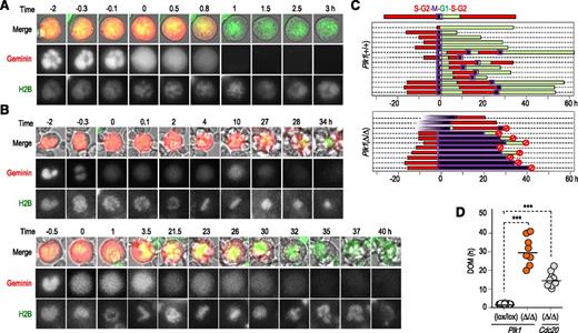
To better follow megakaryocyte polyploidization, we monitored megakaryocyte maturation using time-lapse microscopy in Lin‒ bone marrow cells after treatment with TPO. Bone marrow progenitors were transduced with lentiviral vectors expressing GFP-tagged histone H2B and mCherry fluorescent protein-tagged geminin.25 Cell-cycle progression in control megakaryocytes can be monitored by levels of geminin, which is low in G1 and high in S-phase G2-M, and the pattern of histone that allows following chromosome condensation in mitosis (Figure 3A). Because geminin is exclusively nuclear during interphase, endomitosis can also be detected by pancellular localization of geminin in these cells as a consequence of nuclear envelope breakdown.12 In the absence of Plk1, cells entered normally into mitosis (ie, chromosomes condensed and geminin signal was pancellular because of the absence of a nuclear envelope). However, geminin signal remained high for more than 20 hours during mitosis (chromosomes condensed and pancellular signal of geminin) until cells died (Figure 3B). In a few cases, Plk1(Δ/Δ) megakaryocytes were able to exit from mitosis, but they usually died in the following interphase before they were able to start a new cycle of geminin expression (Figure 3B-C). Whether this death is a direct consequence of Plk1 ablation or is partially due to the intense manipulation of these cultured megakaryocytes is not clear at present. On average, Plk1-deficient megakaryocytes spent about 30 hours in mitosis, whereas the duration of mitosis in normal megakaryocytes was 57 ± 14 minutes (Figure 3D). Interestingly, this arrest in mitosis was even longer than the one obtained after genetic ablation of Cdc20, a model in which mitotic exit is prevented leading to metaphase arrest for long periods until they die.12,26
Defective mitotic exit in Plk1-deficient megakaryocytes. (A) Representative mitotic progression and exit in wild-type megakaryocytes. Cells were transduced with lentiviral vectors expressing GFP-histone H2B (green) and mCherry-Geminin (red). Mitotic entry (time 0) can be monitored by chromosome condensation and pancellular staining of geminin. Mitotic exit (1 hour) is monitored by chromosome decondensation, reformation of nuclear lobules, and geminin degradation. (B) Two independent examples of defective mitotic progression in Plk1-deficient megakaryocytes. Mitotic entry (chromosome condensation and pancellular staining of geminin) is shown at time 0 (third frame in the top cell and second frame in the bottom cell). Geminin is slowly degraded 27 to 32 hours after mitotic entry, resulting in cell death (top cell) or mitotic exit (bottom) after approximately 37 hours. (C) Representation of cell fate in wild-type and Plk1(Δ/Δ) megakaryocytes. Every row represents a single cell. Geminin high phases are shown in red, whereas geminin low phases are in green. Endomitosis is indicated by a purple box and cell death is indicated by a red circle. Mitotic entry (condensation of chromatin and pancellular localization of geminin) is normalized as time 0. (D) Duration of mitosis (DOM; in hours) in Plk1-deficient or control megakaryocytes. Cdc20-deficient megakaryocytes12 are used as a control. ***P < .001; Student t test.
Defective mitotic exit in Plk1-deficient megakaryocytes. (A) Representative mitotic progression and exit in wild-type megakaryocytes. Cells were transduced with lentiviral vectors expressing GFP-histone H2B (green) and mCherry-Geminin (red). Mitotic entry (time 0) can be monitored by chromosome condensation and pancellular staining of geminin. Mitotic exit (1 hour) is monitored by chromosome decondensation, reformation of nuclear lobules, and geminin degradation. (B) Two independent examples of defective mitotic progression in Plk1-deficient megakaryocytes. Mitotic entry (chromosome condensation and pancellular staining of geminin) is shown at time 0 (third frame in the top cell and second frame in the bottom cell). Geminin is slowly degraded 27 to 32 hours after mitotic entry, resulting in cell death (top cell) or mitotic exit (bottom) after approximately 37 hours. (C) Representation of cell fate in wild-type and Plk1(Δ/Δ) megakaryocytes. Every row represents a single cell. Geminin high phases are shown in red, whereas geminin low phases are in green. Endomitosis is indicated by a purple box and cell death is indicated by a red circle. Mitotic entry (condensation of chromatin and pancellular localization of geminin) is normalized as time 0. (D) Duration of mitosis (DOM; in hours) in Plk1-deficient or control megakaryocytes. Cdc20-deficient megakaryocytes12 are used as a control. ***P < .001; Student t test.
Lack of Plk1 results in defective centrosome maturation and abnormal spindle formation in megakaryocytes
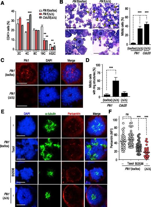
Three days after stimulation with TPO, Plk1(Δ/Δ) CD41+ cells displayed reduced polyploidization (Figure 4A) similar to what was observed in bone marrow cells (Figure 2A). In addition, the percentage of CD41+ cells was enhanced both in Plk1(Δ/Δ) and Cdc20(Δ/Δ) cultures, mainly from the increase in low-complexity CD41+ cells (supplemental Figure 3). These defects were also accompanied by a significant accumulation of mitotic cells as detected by CD41 (not shown) and Giemsa staining (Figure 4B). As in other mitotic cells, Plk1 displayed a diffused localization in control megakaryocytes with some concentration at kinetochores and strong centrosomal signal (Figure 4C). Plk1 immunoreactivity was lost in Plk1(Δ/Δ) megakaryocytes that were characterized by abnormal distribution of mitotic chromosomes frequently forming ringlike structures (Figure 4C-D). These structures were likely a consequence of defective centrosomal maturation and the presence of collapsed spindle poles (Figure 4E). In fact, the staining for pericentrin at centrosomes, a marker of mature organelles, was significantly reduced in Plk1-deficient megakaryocytes (Figure 4E-F). In some cases, pericentrin signals were concentrated in a reduced area, forming a single spindle pole, whereas in other cases, microtubule asters emanated from weak or absent pericentrin marks (supplemental Figure 5). The number of spindle poles was also reduced, indicating lack of centrosome separation or centrosome collapse into a single or a few poles around which chromosomes organized into ring structures (Figure 4E; supplemental Figure 5). As a comparison, mitotic arrest induced by Taxol did not result in decreased pericentrin staining or reduced number of poles (Figure 4E-F). Transient inhibition of Plk1 with the small-molecule compound BI2536 during 12 hours in control cells resulted in similar phenotypes to those observed in the genetic model (Figure 4E-F), suggesting that these defects were a consequence of specific loss of Plk1 activity in megakaryocytes.
Lack of Plk1 results in defective centrosome maturation and spindle formation in mitotic megakaryocytes. (A) Reduced polyploidization in Lin‒ bone marrow cells 3 days after stimulation with TPO. Data are mean ± SD (n = 4 mice per genotype). *P <0.05; **P < .01; ***P < .001; Student t test. (B) Representative images and quantification of mitotic cells (arrowheads) in these cultures 3 days after stimulation with TPO. Scale bars, 20 μm (top) and 50 μm (bottom). Data are mean ± SD. At least 250 cells per genotype were scored. ***P < .001; Student t test. (C) Immunodetection of Plk1 (red) in Plk1(lox/lox) and Plk1(Δ/Δ) stimulated in vitro with TPO. DAPI (blue) was used to stain DNA. Scale bars, 10 μm. (D) Quantification of ring structures in the mitotic cells in these cultures. Data are mean ± SD. n = 50 (control) or 250 (Plk1- or Cdc20-null) mitotic cells per condition. ***P < .001; Student t test. (E) Immunodetection of α-tubulin (green) and pericentrin (red) in Plk1-null or control cells treated with the indicated compounds. DAPI (blue) was used to stain DNA. Scale bars, 10 μm. (F) Quantification of pericentrin median fluorescence intensity (MFI) in these cultures. Horizontal bars indicate averages. ns, not significant; ***P < .001; Student t test.
Lack of Plk1 results in defective centrosome maturation and spindle formation in mitotic megakaryocytes. (A) Reduced polyploidization in Lin‒ bone marrow cells 3 days after stimulation with TPO. Data are mean ± SD (n = 4 mice per genotype). *P <0.05; **P < .01; ***P < .001; Student t test. (B) Representative images and quantification of mitotic cells (arrowheads) in these cultures 3 days after stimulation with TPO. Scale bars, 20 μm (top) and 50 μm (bottom). Data are mean ± SD. At least 250 cells per genotype were scored. ***P < .001; Student t test. (C) Immunodetection of Plk1 (red) in Plk1(lox/lox) and Plk1(Δ/Δ) stimulated in vitro with TPO. DAPI (blue) was used to stain DNA. Scale bars, 10 μm. (D) Quantification of ring structures in the mitotic cells in these cultures. Data are mean ± SD. n = 50 (control) or 250 (Plk1- or Cdc20-null) mitotic cells per condition. ***P < .001; Student t test. (E) Immunodetection of α-tubulin (green) and pericentrin (red) in Plk1-null or control cells treated with the indicated compounds. DAPI (blue) was used to stain DNA. Scale bars, 10 μm. (F) Quantification of pericentrin median fluorescence intensity (MFI) in these cultures. Horizontal bars indicate averages. ns, not significant; ***P < .001; Student t test.
Lack or inhibition of Plk1 results in a SAC-dependent mitotic arrest
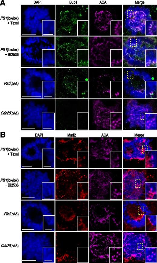
The defects in centrosome maturation and spindle structure suggested that mitotic arrest in Plk1-deficient megakaryocytes could be a consequence of an active SAC. This mitotic checkpoint has been shown to be present in megakaryocytes using Cdc20 as an indirect readout,27 although its relevance has not been established given the lack of genome segregation into separate cells. We therefore monitored the presence of 2 bona-fide SAC proteins, Bub1 and Mad2, in Plk1-deficient megakaryocytes or control cultures. Plk1(Δ/Δ) megakaryocytes or Plk1(lox/lox) cells treated with the Plk1 inhibitor BI2536 displayed a significant accumulation of the SAC components Bub1 (Figure 5A) and Mad2 (Figure 5B) at the kinetochores as detected using ACAs, similarly to control cells treated with Taxol, suggesting the presence of an active SAC. As a negative control, we used Cdc20-deficient megakaryocytes, in which the mitotic arrest is known to occur once the SAC has been satisfied and it is therefore SAC-independent.26
Accumulation of SAC proteins in the kinetochores of Plk1-deficient megakaryocytes. (A) Immunodetection of Bub1 (green) and centromeres (ACAs, magenta) in Plk1(Δ/Δ) cells or control cells treated with Taxol or the Plk1 inhibitor BI2536. Cdc20-deficient cells were used as a control of SAC-independent mitotic arrest. DAPI (blue) was used to stain DNA. (B) Immunodetection of Mad2 (red) and centromeres (ACAs, magenta) in Plk1(Δ/Δ) cells or control cells treated with Taxol or the Plk1 inhibitor BI2536. Cdc20-deficient cells were used as a control of SAC-independent mitotic arrest. DAPI (blue) was used to stain DNA. Scale bars, 10 μm (insets, 2 μm).
Accumulation of SAC proteins in the kinetochores of Plk1-deficient megakaryocytes. (A) Immunodetection of Bub1 (green) and centromeres (ACAs, magenta) in Plk1(Δ/Δ) cells or control cells treated with Taxol or the Plk1 inhibitor BI2536. Cdc20-deficient cells were used as a control of SAC-independent mitotic arrest. DAPI (blue) was used to stain DNA. (B) Immunodetection of Mad2 (red) and centromeres (ACAs, magenta) in Plk1(Δ/Δ) cells or control cells treated with Taxol or the Plk1 inhibitor BI2536. Cdc20-deficient cells were used as a control of SAC-independent mitotic arrest. DAPI (blue) was used to stain DNA. Scale bars, 10 μm (insets, 2 μm).
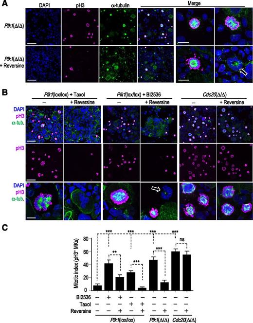
To finally analyze whether this checkpoint was responsible for the mitotic arrest in cells with defective Plk1 function, we use reversine, a small-molecule inhibitor of the SAC kinase Mps1.24 Reversine was able to significantly rescue the accumulation of mitotic cells in Plk1(Δ/Δ) cultures as well as in Plk1(lox/lox) cells treated with BI2536 or Taxol (Figure 6A-C; supplemental Figure 6). In Plk1-deficient cells or in the presence of BI2536, reversine induced mitotic exit, giving rise to cells with interphasic ring nuclei or in which chromosomes formed multiple curved nuclei around the collapsed spindles (Figure 6A-B). Reversine, however, had no effect in the mitotic arrest imposed by the lack of Cdc20 (Figure 6B-C), a defect known to be SAC-independent.26 All together, these observations support the notion that lack of Plk1 prevents mitotic progression and megakaryocyte polyploidization by triggering a SAC-dependent response.
Lack of Plk1 results in a SAC-dependent mitotic arrest. (A) Representative images of Plk1(Δ/Δ) megakaryocytes in the absence or presence of the SAC inhibitor reversine. Spindles are shown by α-tubulin staining (green), whereas pH3 is shown in magenta. DAPI (blue) was used to stain DNA. Scale bars, 50 μm (or 20 μm in the 2 right columns). (B) Representative images of the indicated cultures in the absence or presence of reversine. Spindles are shown by α-tubulin staining (green), whereas pH3 is shown in magenta and DAPI (blue) was used to stain DNA. Scale bars, 50 μm (or 20 μm in the bottom row). (A-B) Arrows indicate curved nuclei around a single tubulin aster. (C) Mitotic index, as determined by detection of pH3, in Plk1- or Cdc20-deficient cells or control cells treated with Taxol or BI2536 in the absence or presence of the Mps1 inhibitor reversine. Data are mean ± SD. n = 50 cells per condition. **P < .01; ***P < .001; Student t test.
Lack of Plk1 results in a SAC-dependent mitotic arrest. (A) Representative images of Plk1(Δ/Δ) megakaryocytes in the absence or presence of the SAC inhibitor reversine. Spindles are shown by α-tubulin staining (green), whereas pH3 is shown in magenta. DAPI (blue) was used to stain DNA. Scale bars, 50 μm (or 20 μm in the 2 right columns). (B) Representative images of the indicated cultures in the absence or presence of reversine. Spindles are shown by α-tubulin staining (green), whereas pH3 is shown in magenta and DAPI (blue) was used to stain DNA. Scale bars, 50 μm (or 20 μm in the bottom row). (A-B) Arrows indicate curved nuclei around a single tubulin aster. (C) Mitotic index, as determined by detection of pH3, in Plk1- or Cdc20-deficient cells or control cells treated with Taxol or BI2536 in the absence or presence of the Mps1 inhibitor reversine. Data are mean ± SD. n = 50 cells per condition. **P < .01; ***P < .001; Student t test.
Discussion
The intrinsic differences between mitotic and endomitotic cell cycles have raised the possibility of using mitotic kinase inhibitors for treating hematological malignancies such as pro-megakaryocytic leukemias.22 Many mitotic kinases such as Aurora A, Aurora B, Cdk1, or Plk1 are essential for the mitotic cell cycles and their genetic ablation results in lack of cell division in the embryo, most somatic cells, and tumor cells.13,15,17,28-30 Yet, inhibition or genetic ablation of Aurora A or Aurora B does not perturb megakaryocyte polyploidization or maturation.9,11-13 In addition, Cdk1 deficiency results in an alternative polyploidization mechanism based on endocycling that has no major effect in platelet production.12 In this article, we report that, contrary to these other mitotic kinases, Plk1 is essential for megakaryocyte polyploidization and its ablation results in thrombocytopenia.
Endomitotic megakaryocytes are characterized by the presence of multipolar spindles with an asymmetrical distribution of chromosomes. Early studies in cell lines suggested that megakaryocytes can polyploidize in the presence of antimicrotubule poisons, suggesting the irrelevance of the SAC in these cells.31 More recent data, however, showed that nocodazole arrests primary megakaryocytes in the presence of Cdc20 signal at the kinetochores, indicating that the SAC may be activated in these cells in the presence of microtubule poisons.27,32,33 Sister chromatid separation seems to occur asymmetrically toward the different poles, and chromosomes cluster into a ring around each aster, suggesting that each chromosome ring may give rise to 1 lobe of the megakaryocytic nucleus.27 The asymmetric segregation of chromosomes toward the multiple poles suggested that the SAC is permissive for the complex attachment of chromosomes to multipolar spindles in megakaryocytes.27 That situation is altered in Plk1-deficient megakaryocytes, likely as a consequence of the formation of monopolar spindles that prevent proper attachment of chromosomes. In the absence of Plk1, all chromosomes are forming a single or a few rings around the only or the few poles present, in agreement with the presence of megakaryocytes with a single or fewer lobules in vivo (see model in supplemental Figure 5). These defects in spindle formation likely lead to a checkpoint-dependent mitotic arrest that, in most cases, triggers cell death, showing that megakaryocytes can also monitor defects in spindle assembly using a functional SAC. The complexity of endomitosis likely requires multiple other pathways, in addition to the Plk1-SAC axis, to generate functional polyploid megakaryocytes with multilobulated nuclei. Yet, the similarities and differences between mitotic and endomitotic cell cycles remain mostly unknown.
The SAC is a complex network that inhibits the APC/C-Cdc20 activity, thus preventing securin and cyclin B degradation and delaying mitotic exit in the presence of unattached kinetochores.34 Yet, cells can be able to exit from mitosis even in the presence of an unsatisfied SAC because of a process known as mitotic slippage.35 During this process, cyclin B1 is slowly degraded as a consequence of weak APC/C-Cdc20 activity and, in fact, mitotic slippage does not occur in Cdc20-null cells and these cells arrest in mitosis until they die.26 That Plk1-deficient megakaryocytes arrest in mitosis for longer periods than Cdc20-deficient megakaryocytes before they die (Figure 3) suggests that Cdc20 and Plk1 may have separate implications in the mechanisms that modulate cell death in mitosis. Indeed, Cdc20 is known to suppress apoptosis through targeting Bim for degradation, suggesting increased mitotic apoptosis in the absence of this APC/C cofactor.36 It has also been recently reported that Plk1-dependent phosphorylation of caspase-8 may trigger the extrinsic apoptotic pathway during mitosis.37 However, treatment of Cdc20-null cells with Plk1 inhibitors does not prolong survival during mitotic arrest (data not shown), and mitotic cell death has been proposed to be mostly mediated by the intrinsic apoptotic pathway.38 Whether Plk1 may directly modulate cell death in megakaryocytes is unclear. Yet, thrombocytopenia is less dramatic in Plk1- compared with Cdc20-mutant models, likely as a consequence of the ability of some Plk1 megakaryocytes to exit from the mitotic arrest and reach higher ploidy levels.
Owing to the essential role of Plk1 in the control of the cell cycle, Plk1 is considered an attractive cancer target with some small-molecule inhibitors showing encouraging results in clinical trials.19,20 Volasertib (a BI2536 derivative also known as BI6727) has shown a significant effect in acute myeloid leukemia patients,39 an effect that recently received a Breakthrough Therapy designation by the Food and Drug Administration, promoting its evaluation in phase 3 clinical trials. Volasertib has also been tested in clinical trials in solid malignancies, in which the most common dose-limiting effects are thrombocytopenia and neutropenia.40,41 These problems may be due to specific effects in progenitors cells or that Plk1 inhibitors can inhibit other kinases at specific doses.42,43 However, our conditional knockout model suggests that Plk1 inhibition actually leads to defective polyploidization and cell death in megakaryocytes, a result with implications in the design of therapeutic protocols targeting this mitotic kinase.
The online version of this article contains a data supplement.
The publication costs of this article were defrayed in part by page charge payment. Therefore, and solely to indicate this fact, this article is hereby marked “advertisement” in accordance with 18 USC section 1734.
Acknowledgments
The authors are indebted to V. Andrés (Centro Nacional de Investigaciones Cardiovasculares, Madrid, Spain) and A. Miyawaki (RIKEN, Saitama, Japan) for reagents, Lola Martínez and Diego Megías (Centro Nacional de Investigaciones Oncológicas [CNIO]) for help with cytometry and microscopy analysis, Fernando Escobar and Begoña Pou (Centro de Investigaciones Biológicas, Madrid, Spain) for help in the preparation and analysis of electron microscopy samples, members of the Comparative Pathology and Transgenic Units of the CNIO for excellent technical support, and Sara Leceta and Sheila Rueda for help with animal management.
This work was supported by a fellowship from the Foundation La Caixa (M.T.), and the Cell Division and Cancer group of the CNIO was funded by the Ministry of Economy and Competitiveness (SAF2012-38215), Consolider-Ingenio 2010 Programme (SAF2014-57791-REDC), Red Temática CellSYS (BFU2014-52125-REDT), Comunidad de Madrid (OncoCycle Programme, S2010/BMD-2470), Worldwide Cancer Research (WCR #15-0278), and the MitoSys project (HEALTH-F5-2010-241548, European Union Seventh Framework Programme).
Authorship
Contribution: M.T. generated the mouse models, performed the experiments, and analyzed the data; D.P., M.S.-R., G.d.C., and M. Maroto contributed to the cellular studies; P.W. generated the Plk1 conditional allele; and M. Malumbres designed and supervised the project, analyzed the data, and wrote the manuscript.
Conflict-of-interest disclosure: The authors declare no competing financial interests.
Correspondence: Marcos Malumbres, Centro Nacional de Investigaciones Oncológicas (CNIO), Melchor Fernández Almagro 3, E-28029 Madrid, Spain; e-mail: malumbres@cnio.es.

This feature is available to Subscribers Only
Sign In or Create an Account Close Modal