Key Points
Establishing HSC transplantation and assay methods for the axolotl.
Axolotl sites of hematopoiesis are the spleen and liver.
Abstract
Hematopoietic stem cell (HSC)-derived cells are involved in wound healing responses throughout the body. Unfortunately for mammals, wound repair typically results in scarring and nonfunctional reparation. Among vertebrates, none display such an extensive ability for adult regeneration as urodele amphibians, including 1 of the more popular models: the axolotl. However, a lack of knowledge of axolotl hematopoiesis hinders the use of this animal for the study of hematopoietic cells in scar-free wound healing and tissue regeneration. We used white and cytomegalovirus:green fluorescent protein+ transgenic white axolotl strains to map sites of hematopoiesis and develop hematopoietic cell transplant methodology. We also established a fluorescence-activated cell sorter enrichment technique for major blood lineages and colony-forming unit assays for hematopoietic progenitors. The liver and spleen are both active sites of hematopoiesis in adult axolotls and contain transplantable HSCs capable of long-term multilineage blood reconstitution. As in zebrafish, use of the white axolotl mutant allows direct visualization of homing, engraftment, and hematopoiesis in real time. Donor-derived hematopoiesis occurred for >2 years in recipients generating stable hematopoietic chimeras. Organ segregation, made possible by embryonic microsurgeries wherein halves of 2 differently colored embryos were joined, indicate that the spleen is the definitive site of adult hematopoiesis.
Introduction
In mammals, the ability to regenerate limbs and organs is progressively lost during ontogeny and correlates closely with maturation of immune competence. Research in scar-free healing, primarily observed in model systems with dysfunctional neutrophils and macrophages, has led to the hypothesis that the immune system dictates the balance between scarring and regeneration.1,2 Unfortunately, presently available genetic models of vertebrate wound healing, such as the African spiny mouse (Acomys), tend to lack significant regenerative abilities.3 Thus, although the role of hematopoietic stem cell (HSC)-derived blood cells in wound healing via inflammation and paracrine regulation is well understood during fibrotic healing, the same cannot be said for a scar-free regenerative response.4,5
Among vertebrates, urodele amphibians, such as the axolotl (Ambystoma mexicanum), display a unique and extensive ability for adult regeneration. Axolotls can replace a wide variety of tissues and complex structures including muscle, cartilage, skin, spinal cord, brain, heart, jaw, and limbs.6 The axolotl’s nearest relatives, anuran frogs and reptiles, lose the ability to reform tissues after metamorphosis or show limited tail regeneration. This suggests that adult regeneration may be an acquired trait in axolotls that might be inducible in mammals if the required genetic pathways are mapped. Recent advances in production of transgenic axolotls,6-10 complete mapping of the axolotl transcriptome,11 and production of gene expression arrays12-16 finally allow molecular mapping of regeneration pathways. Additionally, given the extensive conservation of synteny between axolotls and humans, this amphibian may be a powerful genetic model to study hematopoietic cell function in scar-free wound healing and tissue regeneration.17
However, 1 major challenge in using axolotls is a limited knowledge of their hematopoiesis. Axolotls produce similar blood lineages as mammals with the exception of persistent orthochromatic normoblasts in adults.18,19 Yet to be defined are fundamental questions such as sites of hematopoiesis and the HSC niche. Therefore, in this study, we define the primary and secondary sites of hematopoiesis in both larval and adult axolotls. Furthermore, we developed the classic tools of hematology to study axolotl hematopoiesis including hematopoietic cell transplantations (HCTs), colony-forming unit (CFU) assays, and methods to map axolotl hematopoiesis. Finally, we have taken advantage of axolotl embryo manipulations to generate stable hematopoietic chimeras (green fluorescent protein [GFP]+ blood in a white recipient) in nonirradiated models with directly visible immune cells similar to the zebrafish.
Materials and methods
Axolotls
White mutant (d/d), GFP+, or nucCherryRed+ cytomegalovirus (CMV):β-actin promoter-driven transgenic axolotls and embryos were purchased from the Ambystoma Genetic Stock Center (AGSC) or bred in-house from AGSC founder animals. Animals were staged as previously described20 and maintained in Holtfreter’s solution.21 This study was approved under the University of Florida Institutional Animal Care and Use Committee protocol 201202645.
Irradiation
Adult axolotls were anesthetized by submersion in 0.1% tricaine-methanesulfonate (Sigma-Aldrich), pH 7.4, and a 900- to 1000-cGy dose of irradiation22 was given in a 137Cs source irradiator with or without lead shielding used to protect the gills, or a 1000- to 1200-cGy dose of γ-irradiation was specifically targeted to the spleen and liver region via a six Mev linear accelerator.
Cell collection
As a general note, axolotl cells require a different osmotic pressure than mammalian cells, thus requiring standard buffers/media be diluted to 75% normal strength with water prior to use. Axolotl cells were maintained in either 0.60× L-15 media, 5% fetal bovine serum (FBS), 1× penicillin/streptomycin, and 1× insulin-transferrin or 0.75× phosphate-buffered saline (PBS) (referred to as axolotl PBS [APBS]). Cells were kept at 18°C to 20°C in ambient air. Axolotls were anesthetized, tissues were collected, and single cell suspensions were made by maceration in APBS and 5% FBS. Axolotl blood was collected into 10 mM EDTA in APBS to prevent coagulation. Ficoll-Paque Plus (GE) density gradients were performed as described elsewhere23 for lymphoblastic “buffy” coat and erythrocyte enrichment.
Flow cytometry
Single-cell suspensions of liver and spleen cells from GFP+ axolotls in APBS with 5% FBS were stained with 7-amino-actinomycin D (BD Biosciences) to exclude dead cells and debris. Analysis and sorting was accomplished on a fluorescence-activated cell sorter (FACS) Aria flow cytometer (BD Biosciences) based on 7-amino-actinomycin D exclusion, GFP fluorescence, side scatter (SSC), and forward scatter (FSC) for cell size and granularity.
Histology
Tissues were dissected and fixed in 4% paraformaldehyde at 4°C, washed with PBS, and transferred to 20% sucrose in PBS for 3 days at 4°C. Tissues were embedded in Optimal Cutting Temperature Compound (Sakura Finctek) and stored at −20°C. Sections were cut to a thickness of 5 µm and stained with hematoxylin and eosin (H&E). Transferase-mediated deoxyuridine triphosphate nick-end labeling (TUNEL) staining was performed using the In Situ Cell Death Detection Kit, TMR red (Roche), in accordance with the manufacturer’s instructions. Cytospins were made with 5 × 10⁴ to 1 × 10⁵ cells. Samples were spun at 800 rpm for 3 minutes onto glass slides. Prepared slides were stained with Protocol HEMA 3 stain set (Fisher Scientific), Wright-Giemsa, Alcian blue, or α-naphthyl butyrate esterase. Antibodies were mouse monoclonal F4/80 IgG (Serotec; MCAP497) 1:50, rabbit polyclonals CD79a IgG (Abcam; AB5691) 1:100, and CD2 IgG (AB37212; Abcam) 1:50. Microscopy of tissue sections and cytospins was performed on a Leica DM5500B microscope using a Hamamatsu digital camera model C7780.
Phagocyte assay
pHrodo Red Escherichia coli BioParticles (Molecular Probes) were mixed with whole blood and incubated at room temperature for 1 to 3 hours. Erythrocytes were separated by Ficoll-Paque density gradient, and all other fractions were cytospun onto glass slides for visualization.
Hematopoietic cell transplantation
Lethally irradiated (950 cGy)22 white adult animals were anesthetized and received a minimum of 1 × 10⁴ whole spleen or liver cells intravenously through a 26-gauge needle in a maximum volume of 300 µL. Microinjections into nonirradiated embryos (stages 25 to hatching) and larvae (3 months old) were performed using borosilicate glass capillary needles (1-mm outer diameter, no filament; World Precision Instruments) made using a micropipette puller. Procedures were done via micromanipulator and a screw-actuated air/oil microinjector; 1 × 10⁴ to 5 × 10⁵ cells were injected intracardially into tricaine anesthetized animals. Axolotls were imaged on a Leica MZ16FA microscope using a Hamamatsu digital camera model C7780 and Volocity Imaging software (Perkin Elmer).
Fused 2-color chimeras
Embryos at stages 14 to 20 were dejelled and washed in fresh 100% Steinberg’s solution24 with a pH of 7.4 containing 1% antibiotic-antimycotic and 0.0025% gentamycin (25 mg/L). Under a dissecting microscope, embryos of each color were cut transversely in half with microsurgical scissors, and the anterior end of one was matched with the posterior end of the other. Paired halves were moved into depressions made in agar with neural folds touching. Embryos were left undisturbed for 96 hours at 20°C, transferred into fresh 100% Steinberg’s solution for another 7 days, and then moved to 40% Holtfreter’s solution.
CFU assays
Axolotl cellular responses to mammalian colony stimulating factors (CSFs) are unknown. Therefore, we produced axolotl pokeweed mitogen-stimulated spleen cell-conditioned media (PWM-SCM) to serve as a source of axolotl CSFs for CFU assays. Axolotl spleen cells were suspended in 60% L-15 media, 10% PBS, penicillin/streptomycin, insulin-transferrin-selenium, and 1% pokeweed mitogen solution (1 mg/mL), pH 6.4, at a cell concentration of 1 × 10⁶ to 2 × 10⁶ cells/mL, and allowed to condition medium for 7 days at 18°C. Conditioned media were collected by centrifugation, filtered through a 0.45-µm filter, and stored at −80°C.
Single-cell suspensions of spleen and liver cells were suspended at a final concentration of 5 × 10⁴ cells/mL in 3 mL of 2% methylcellulose (Methocel), 50% PWM-SCM, and 0.60× L-15 media, pH 6.4, in 35-mm Petri plates. Human erythropoietin was added at 1 U/mL. Cultures were incubated up to 5 weeks at 18°C in ambient air.
Polymerase chain reaction
Genomic DNA was isolated from whole blood or Ficoll-Paque Plus density gradient-purified erythrocyte fractions and prepared as described elsewhere.23 Primers were designed to amplify a 173-bp region within the GFP gene. The forward primer was AAGTTCATCTGCACCACCG, and the reverse primer was TCCTTGAAGAAGATGGTGCG. Reactions (25 µL) were prepared with the addition of 4% dimethylsulfoxide, and the thermal cycler was programed as described elsewhere.25
Statistical analysis
Microsoft Excel 2013 was used to perform 2-tailed Student t tests. P < .05 was considered significant.
Results
Defining sites of hematopoiesis in the axolotl
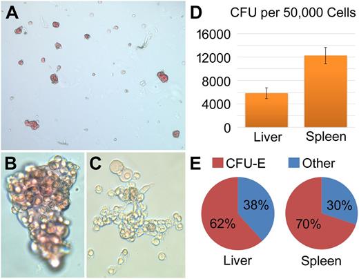
Previous studies of the axolotl immune system noted that the liver and spleen both contain significant numbers of hematopoietic cells, whereas the bone marrow appears to be nonhematopoietic.22,26,27 To confirm which organs in adult axolotls contain hematopoietic progenitor cells (HPCs), we developed an axolotl-specific CFU assay based on early mouse CFU assays using PWM-SCM as a source of CSFs. We tested the addition of a variety of mammalian growth factors routinely used for serum-free CFU assays. Only erythropoietin (EPO) induced formation of additional colonies. Axolotl cells are notoriously difficult to grow in tissue culture. To date, only a single cell line (AL-1 fibroblasts)28 has been established from axolotls. Primary cultures of axolotl cells usually fail after only a few passages. Colonies generated from axolotl (N = 5 each organ) spleen and liver cells in response to axolotl PWM-SCM/EPO were similar in morphology to classically defined erythroid burst-forming unit and erythroid CFU (CFU-E) (Figure 1A-B). Given how little is known about hematopoiesis/hematopoietic development in axolotls, we use the term CFU-E to describe all erythroid-only colonies in this study. The second major colony type present resembled granulocyte, erythrocyte, monocyte, megakaryocyte CFU (CFU-GEMM) colonies (Figure 1C) containing a mixture of myeloid, megakaryocyte, and erythroid lineage cells. After culture initiation, colonies were first visible by 4 days and peaked at 10 days. Axolotl spleens generated twice the number of total colonies than livers (12 253 vs 5825, P = .0009; Figure 1D). Axolotl spleen and liver generated similar proportions of CFU-E (70% vs 62%, P = .4) and CFU-GEMM (30% vs 38%, P = .4) colonies (Figure 1E). We also tested cells from all other major organs including bone marrow, thymus, and kidneys for CFU activity and found none. Therefore, we concluded that spleen and liver are the active sites of hematopoiesis in the adult axolotl.
HPC activity from axolotl spleen and liver. (A) Representative view (×10) of spleen CFU at 10 days. (B) Magnification (×40) of a large CFU-E colony. (C) Magnification (×40) of a non-CFU-E colony. (D) Quantitation of CFUs in the liver and spleen reveals that the spleen contains more than twice as many CFUs as the liver (12 253 vs 5825, P = .0009; N = 3). Graphs show mean ± standard error of the mean of ≥3 independent experiments. (E) The proportion of CFU-E to other CFU colonies (P = .4). CFU assays were imaged on a Leica DMIRB inverted microscope using a SPOT Flex digital camera.
HPC activity from axolotl spleen and liver. (A) Representative view (×10) of spleen CFU at 10 days. (B) Magnification (×40) of a large CFU-E colony. (C) Magnification (×40) of a non-CFU-E colony. (D) Quantitation of CFUs in the liver and spleen reveals that the spleen contains more than twice as many CFUs as the liver (12 253 vs 5825, P = .0009; N = 3). Graphs show mean ± standard error of the mean of ≥3 independent experiments. (E) The proportion of CFU-E to other CFU colonies (P = .4). CFU assays were imaged on a Leica DMIRB inverted microscope using a SPOT Flex digital camera.
One limitation to hematologic analysis in axolotl is a lack of cell-specific antibodies that work for flow cytometry. For transplant studies, we use GFP+ animals as HSC donors, thereby permitting detection of erythroid, myeloid, and lymphoblastic cells via a combination of FSC, SSC, and GFP. Analysis of peripheral blood, spleen, and liver confirmed that these organs contain multiple hematopoietic lineages (Figure 2).22 Spleens are mostly composed of hematopoietic cells, whereas livers have a thin outer layer of predominantly hematopoietic cells with rarer hematopoietic cells found in the inner hepatocyte dominated layers. Erythrocytes, mature myeloid cells, and lymphoblasts were sorted based on FSC/SSC/GFP properties, and their enriched populations were confirmed by cytospin, H&E, and Wright-Giemsa staining (Figure 2A-D). The lymphoblastic population is likely to contain not only B and T lymphocytes, but monocytes and HPCs depending on tissue source. The mature myeloid cell population contained Alcian blue-positive mast cells, α-naphthyl butyrate esterase-expressing macrophages, and myeloperoxidase-positive neutrophils18,29-33 (Figure 2D).
Identification of major hematopoietic cell lineages from the liver and spleen by FACS light-scatter characteristics, fluorescence, and staining. FACS and H&E staining are presented for (A) axolotl blood, (B) liver, and (C) spleen. The CMV:GFP transgene expresses in all tissue types, but not in every cell. Depending on cell lineage, GFP expression can vary from 10% to 90% of individual cells. This is similar to the variable transgene expression seen in the equivalent transgenic mouse.31 All erythrocytes are GFP− (red). Mature myeloid cell enriched populations are GFP+SSChi (blue). Lymphocyte enriched populations are GFP+SSClo (blue). The liver contains almost even numbers of myeloid cells and lymphoblasts. The spleen contains more lymphoblasts than myeloid cells. Black cells are excluded debris. (D) Cytospins followed by Wright-Giemsa, Alcian blue, α-naphthyl acetate esterase, and myeloperoxidase staining were performed to confirm cell morphology and characteristics. Scale bar: 20 µm (applies to the 8 individual cells stained with Wright-Giemsa). Blood composition was 3.7 ± 2.5% lymphoblastic cells, 1.3 ± 0.5% mature myeloid cells, and 95 ± 1.6% erythrocytes. Following removal of adipocytes, the liver was composed of 3.7 ± 0.6% lymphoblastic cells, 5.3 ± 1.5% mature myeloid cells, and 68.5 ± 10.2% erythrocytes (N = 3; Figure 1A). In comparison, the spleen contained 31.3 ± 12% lymphoblastic cells, 5 ± 0.8% mature myeloid cells, and 60.7 ± 15.5% erythrocytes (N = 3).
Identification of major hematopoietic cell lineages from the liver and spleen by FACS light-scatter characteristics, fluorescence, and staining. FACS and H&E staining are presented for (A) axolotl blood, (B) liver, and (C) spleen. The CMV:GFP transgene expresses in all tissue types, but not in every cell. Depending on cell lineage, GFP expression can vary from 10% to 90% of individual cells. This is similar to the variable transgene expression seen in the equivalent transgenic mouse.31 All erythrocytes are GFP− (red). Mature myeloid cell enriched populations are GFP+SSChi (blue). Lymphocyte enriched populations are GFP+SSClo (blue). The liver contains almost even numbers of myeloid cells and lymphoblasts. The spleen contains more lymphoblasts than myeloid cells. Black cells are excluded debris. (D) Cytospins followed by Wright-Giemsa, Alcian blue, α-naphthyl acetate esterase, and myeloperoxidase staining were performed to confirm cell morphology and characteristics. Scale bar: 20 µm (applies to the 8 individual cells stained with Wright-Giemsa). Blood composition was 3.7 ± 2.5% lymphoblastic cells, 1.3 ± 0.5% mature myeloid cells, and 95 ± 1.6% erythrocytes. Following removal of adipocytes, the liver was composed of 3.7 ± 0.6% lymphoblastic cells, 5.3 ± 1.5% mature myeloid cells, and 68.5 ± 10.2% erythrocytes (N = 3; Figure 1A). In comparison, the spleen contained 31.3 ± 12% lymphoblastic cells, 5 ± 0.8% mature myeloid cells, and 60.7 ± 15.5% erythrocytes (N = 3).
Ablative transplants in the axolotl
Next we developed HCT protocols for axolotl to test for HSC activity and to define its primary niche(s) during axolotl development. Initial transplants adapted our murine ablative bone marrow transplant protocol to the axolotl. White axolotl mutants were used as recipients (Figure 3A) to allow for noninvasive visualization of internal tissues and organs as in zebrafish, but with the added benefits of increased size and visualization persisting throughout adulthood. This trait facilitated monitoring of donor GFP+ cell engraftment after transplant. Cohorts of adult white axolotls received 950-cGy whole body irradiation, with lead shielding of the head to protect the gills from damage, followed by intravenous transplant of test GFP+ donor cell populations. We tested all tissues/organs to avoid bias. Standard transplants received 5 × 10⁶ cells from test organs, or all cells from smaller organs into a single recipient if the yield was lower (ie, bone marrow flushing yielded 1 × 10⁵ cells per donor). From adult donors, only recipients of liver or spleen cells showed a progressive reconstitution with GFP+ blood easily visualized in white recipients (Figure 3B-F). Increasing numbers of GFP+ leukocytes could be seen flowing through blood vessels (Figure 3C; supplemental Videos 1 and 2, available on the Blood Web site) that stabilized after 5 to 6 months. Accumulation of GFP+ cells was visible in what appears to be a lymph-like network beneath the skin (Figure 3B). When adult spleen/liver was used as a donor source, excellent initial reconstitution was observed, but the majority of recipients (>75%) would develop symptoms consistent with graft-versus-host disease (GVHD) by 2 to 5 months after transplant (supplemental Figure 1). Previous studies on axolotl immune function suggested that larvae from 3 to 7 months of age still have immature immune systems.34-36 Therefore, we used larvae in this age range as donors for transplantation. These transplants showed a reduced incident of GVHD (<25%) and robust GFP+ hematopoietic reconstitution. We now use 3- to 7-month-old donor tissue for transplant into gill-shielded, whole body irradiated recipients as current “best practice” for ablative HCT experiments. We have observed up to 20% (1% GFP+ cells in peripheral blood vs 5% average for donors) chimerism of donor cells within the first 3 months. Ficoll-Paque density centrifugation was used to enrich for lymphoblastic cells (buffy coat). Transplantation of 104 GFP+ lymphoblastic spleen cells resulted in equivalent levels of donor cell engraftment as 107 unfractionated GFP+ spleen cells, a roughly 1000-fold enrichment. We concluded that in adult axolotls, both liver and spleen contained HSC populations that are enriched in the lymphoblastic population.
Organs and donor derived GFP+ hematopoietic cells can be visualized directly through the skin in the axolotl. (A) The underside of a wild-type white larval axolotl. The internal organs can be seen clearly through the skin. (B) The head of an irradiated white adult axolotl 1 month after HCT. The iris of the eyes (arrows) are autofluorescent but the nodes are regions of highly concentrated GFP+ donor-derived cells. (C) GFP channel with lighting to visualize GFP+ cells and non-GFP vasculature. Two GFP+ blood cells (white arrowheads) are flowing through vasculature in the tail of a transplanted axolotl in this frame. The orange is white light diffracting off of non-GFP tissues. (D) The foot of a transplanted axolotl with a patch of GFP+ HSPC-derived cells in the skin. Dashed line indicates a previous amputation site (transverse cut) and the section used for E. Dense recruitment of donor-derived cells suggests the potential involvement of blood cells in limb regeneration. (E) DIC image (×20) of the foot sectioned in D. Superior aspect of the inferior portion of the foot. Blue, DAPI staining of cell nuclei; green, donor-derived GFP+ cells. Scale bar: 100 µm. (F) Dissecting microscope view of static donor-derived cells in the skin.
Organs and donor derived GFP+ hematopoietic cells can be visualized directly through the skin in the axolotl. (A) The underside of a wild-type white larval axolotl. The internal organs can be seen clearly through the skin. (B) The head of an irradiated white adult axolotl 1 month after HCT. The iris of the eyes (arrows) are autofluorescent but the nodes are regions of highly concentrated GFP+ donor-derived cells. (C) GFP channel with lighting to visualize GFP+ cells and non-GFP vasculature. Two GFP+ blood cells (white arrowheads) are flowing through vasculature in the tail of a transplanted axolotl in this frame. The orange is white light diffracting off of non-GFP tissues. (D) The foot of a transplanted axolotl with a patch of GFP+ HSPC-derived cells in the skin. Dashed line indicates a previous amputation site (transverse cut) and the section used for E. Dense recruitment of donor-derived cells suggests the potential involvement of blood cells in limb regeneration. (E) DIC image (×20) of the foot sectioned in D. Superior aspect of the inferior portion of the foot. Blue, DAPI staining of cell nuclei; green, donor-derived GFP+ cells. Scale bar: 100 µm. (F) Dissecting microscope view of static donor-derived cells in the skin.
We also used a 6-Mev linear accelerator to provide 1000 cGy of irradiation targeted specifically to the spleen and liver. These cohorts reconstituted well with adult GFP+ spleen and/or liver cells. An example from these cohorts is seen in Figure 3D-E. This animal happened to have a front limb eaten by its tankmates. This limb regenerated with participation of large numbers of donor-derived GFP+ cells during tissue remodeling/regeneration, which is a result consistent with a recent study showing macrophages are required for axolotl limb regeneration.29 Targeted irradiation of liver and spleen without transplant resulted in hematopoietic failure/anemia necessitating euthanasia by 6 weeks, demonstrating that they are the primary hematopoietic organs.
We confirmed the effects of irradiation on liver and spleen by TUNEL staining. Spleens showed a gradual increase in apoptotic cells until nearly every cell appeared TUNEL+ at 7 weeks (supplemental Figure 2A,D,G). The number of apoptotic cells in liver seemed to remain relatively constant and mostly in the periphery (supplemental Figure 2B,E,H). The tail (composed mainly of muscle) served as a radioresistant control and had a small increase in apoptotic cells after irradiation that continued unchanged from 3 to 7 weeks (supplemental Figure 2C,F,I).
Representative histology of HCT recipients (N = 10 animals) shows a distinct peripheral layer in liver composed primarily of lymphoblastic cells (Figure 4A) that was ablated entirely within 1 month of irradiation, whereas parenchymal liver cells showed minimal signs of ablation (Figure 4C). Following irradiation, spleens demonstrated a decrease in lymphoblasts and erythrocytes, but no distinct regions or cell types were differentially affected (Figure 4B,D). Tissues examined at 3 weeks after irradiation and transplant showed widespread engraftment in spleen and liver (Figure 4E-H). Interestingly, engraftment in liver appeared to concentrate in a peripheral hematopoietic layer (Figure 4E,G). Engraftment in spleen was evenly distributed throughout the organ (Figure 4F,H). In 6-week-old livers, GFP+ donor cell engraftment was considerably less than seen in spleen without major preference for the periphery (Figure 4I,K). Engraftment in spleen remained extensive (Figure 4J,L). Overall, there were a noticeably higher proportion of GFP+ cells engrafted in spleen than in liver (Figure 4M-N) at 6 weeks after transplant.
Effects of ablative HCT on the liver and spleen. Characteristic histology analysis of irradiated and or transplanted axolotl. The examples shown are from recipients of both spleen and liver cells, but the results are similar in spleen or liver only transplants. No other donor tissue transplant resulted in GFP+ donor cell engraftment. (A,C) H&E staining (×40) of the liver. Irradiation destroys the peripheral hematopoietic layer of the liver demarcated by the red line and “H”. The black line marks where the hematopoietic layer ends and hepatocytes begin. (B,D) H&E staining (×40) of the spleen. The tissue displays some degradation after irradiation. (E,G) H&E staining (×20) of the liver with matching direct GFP 3 weeks after irradiation and transplant. The hematopoietic peripheral layer is still present but partially ablated and shows more engraftment by donor cells than the rest of the liver. (F,H) H&E staining (×20) of the spleen with matching direct GFP 3 weeks after irradiation and transplant. Donor cells engraft all through the organ without preference as seen in the liver. (I,K) H&E staining (×20) of the liver with matching direct GFP 6 weeks after irradiation and transplant. The hematopoietic layer has been ablated and donor cells show poor engraftment without preference for the periphery. (J,L) H&E staining (×20) of the spleen with matching direct GFP 6 weeks after irradiation and transplant. There is no change in engraftment as seen in the liver. (M-N) Direct GFP section (×5) of the liver and spleen at 4 weeks after irradiation and transplant. Donor cells show some preference for engraftment near the periphery in the liver but none in the spleen and there is substantially more engraftment in the spleen.
Effects of ablative HCT on the liver and spleen. Characteristic histology analysis of irradiated and or transplanted axolotl. The examples shown are from recipients of both spleen and liver cells, but the results are similar in spleen or liver only transplants. No other donor tissue transplant resulted in GFP+ donor cell engraftment. (A,C) H&E staining (×40) of the liver. Irradiation destroys the peripheral hematopoietic layer of the liver demarcated by the red line and “H”. The black line marks where the hematopoietic layer ends and hepatocytes begin. (B,D) H&E staining (×40) of the spleen. The tissue displays some degradation after irradiation. (E,G) H&E staining (×20) of the liver with matching direct GFP 3 weeks after irradiation and transplant. The hematopoietic peripheral layer is still present but partially ablated and shows more engraftment by donor cells than the rest of the liver. (F,H) H&E staining (×20) of the spleen with matching direct GFP 3 weeks after irradiation and transplant. Donor cells engraft all through the organ without preference as seen in the liver. (I,K) H&E staining (×20) of the liver with matching direct GFP 6 weeks after irradiation and transplant. The hematopoietic layer has been ablated and donor cells show poor engraftment without preference for the periphery. (J,L) H&E staining (×20) of the spleen with matching direct GFP 6 weeks after irradiation and transplant. There is no change in engraftment as seen in the liver. (M-N) Direct GFP section (×5) of the liver and spleen at 4 weeks after irradiation and transplant. Donor cells show some preference for engraftment near the periphery in the liver but none in the spleen and there is substantially more engraftment in the spleen.
Collectively, these results indicate that both liver and spleen contain transplantable HSCs and are active sites of hematopoiesis.
Nonablative in utero/larval transplants prevent GVHD
Irradiative conditioning has been shown to impair regeneration of the axolotl.37,38 To avoid irradiation, and generate stable blood chimeras, 2 microsurgical techniques in embryonic and larval axolotls were used: parabiosis and intracardiac microinjection HCT. Parabiosis allows anatomical joining of 2 embryos resulting in a shared circulation. The procedure requires 1 surgery to fuse a GFP+ embryo with a white mutant embryo side by side (supplemental Figure 3A-D), and then another 24 hours later to denervate the GFP+ embryo. Denervation prevents injury as the 2 axolotls develop together. It is then possible to track GFP+ blood in the white axolotl (supplemental Figure 3E-F). This method proved useful for short-term studies, but difficulties with rearing parabiotic axolotls to adulthood compelled us to explore further options.
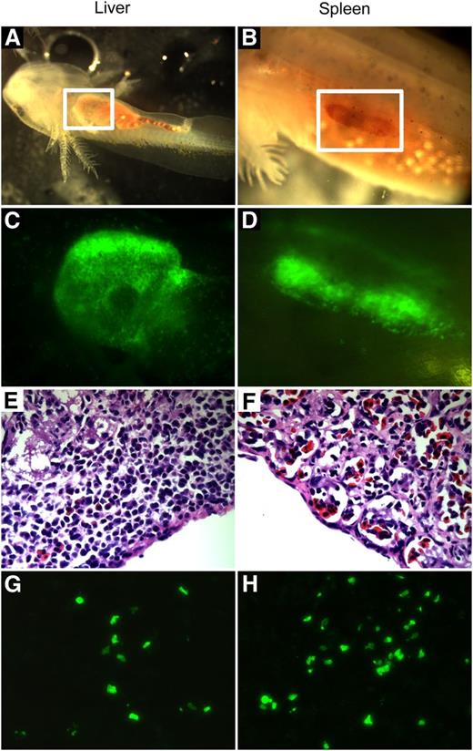
HCT normally requires chemical or irradiative conditioning, and GVHD is expected in unmatched donor-recipient pairs. One exception across a number of species is HCT in the embryonic stages of development. Thus, we sought to induce tolerance by intracardiac microinjection (N = 25) of GFP+ hematopoietic cells into recipient embryos and larvae prior to immune system maturation (up to 3 months of age). In our experiments, 18 of 25 (72%) recipients of whole adult liver or spleen cells showed persistence of GFP+ blood for the life of the animal with varying levels of chimerism (4.0-8.0% in blood). GFP+ cells were directly visualized homing and engrafting in the larval liver (Figure 5A,C) and spleen (Figure 5B,D) as the animals developed irrespective of cell origin. GVHD was never observed in animals that underwent the procedure from stage 36 to 3 months of age. In transplants of 3-month-old larvae, donor engraftment persisted for >2 years in 9 of 10 recipients of ≥1 × 10⁴ whole adult spleen or liver cells (Figure 5E-H) with blood chimerism levels at 4% to 8%. Fewer axolotls showed engraftment, and blood chimerism was undetectable beyond 2 months when less than 1 × 10⁴ spleen cells were injected. In comparison with irradiated chimeras, microinjected axolotls showed fewer GFP+ donor cells in the liver’s peripheral hematopoietic layer (Figure 5E,G). However, spleens showed greater engraftment than livers as previously observed in irradiated chimeras (Figure 5G-H). Together, these results provide conclusive evidence that both liver and spleen contain HSCs capable of long-term durable engraftment.
Donor HSCs engraft in the liver and spleen in microinjected embryos and larvae to create stable chimeras. (A) Anesthetized larva 1 month after microinjection transplant with the liver visible through the skin (white box). (B) Anesthetized 3-month-old larvae 4 days after microinjection transplant with the spleen visible through the skin (white box). (C-D) In vivo GFP channel view of the magnified liver in A and spleen in B shows engraftment of a large number of GFP+ donor cells. (E-F) H&E staining (×40) of the liver and spleen from a 2-year-old microinjected chimera. (G) Section (×40) displaying donor GFP+ cell engraftment in the liver in E. No preference for the peripheral hematopoietic layer is visible. (H) Section (×40) displaying greater donor GFP+ cell engraftment in the spleen in F than in the liver.
Donor HSCs engraft in the liver and spleen in microinjected embryos and larvae to create stable chimeras. (A) Anesthetized larva 1 month after microinjection transplant with the liver visible through the skin (white box). (B) Anesthetized 3-month-old larvae 4 days after microinjection transplant with the spleen visible through the skin (white box). (C-D) In vivo GFP channel view of the magnified liver in A and spleen in B shows engraftment of a large number of GFP+ donor cells. (E-F) H&E staining (×40) of the liver and spleen from a 2-year-old microinjected chimera. (G) Section (×40) displaying donor GFP+ cell engraftment in the liver in E. No preference for the peripheral hematopoietic layer is visible. (H) Section (×40) displaying greater donor GFP+ cell engraftment in the spleen in F than in the liver.
First transplantable HSC arises in the embryonic axolotl liver
To determine at which point in embryonic development transplantable HSCs can be isolated, hematopoietic cells were collected from GFP+ axolotls throughout development. Cells from blood islands (developing at stages 32-38),31 larval livers (developing at stage 39), and larval spleens (developing after hatching) were used as graft sources. No animals receiving blood island cell transplants showed evidence of donor hematopoiesis. However, all animals receiving larval liver cell transplant or larval spleen cell transplant exhibited donor engraftment (data not shown). These results imply that a transplantable HSC first develops in the axolotl liver at stage 39 prior to the formation of the spleen. On formation, the spleen also contains transplantable HSC in the larvae, which is a condition retained through adulthood.
Spleen supports long-term HSC self-renewal in adult axolotl
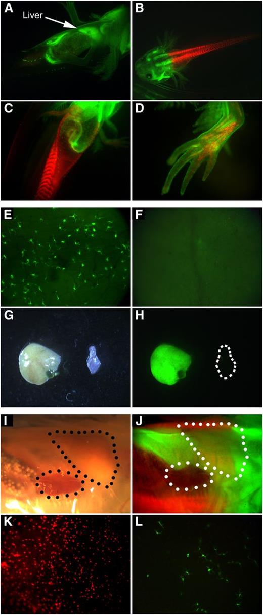
Because both livers and spleens in embryos and adult axolotls contain transplantable HSCs, our next goal was to determine if one or both organs provide a microenvironment capable of supporting long-term HSC (LT-HSC) self-renewal. By bisecting GFP+ embryos and fusing the cephalic portion with the caudal portion of either white or nucCherryRed+ embryos (Figure 6A-D), spleens would develop either the same or a different color than livers facilitating identification of the long-term HSC niche. Nine of 9 (100%) animals showed evidence of GFP+ hematopoiesis for the first 5 months in the juvenile stage of axolotl development (Figure 6E), but as the animals developed into adulthood (>5 months), GFP+ hematopoiesis gradually disappeared by 9 months in some axolotls. In these adult animals, GFP+ cells first disappeared from vasculature and then from skin (Figure 6F). On examination of internal organs, the liver and other organs were found to be GFP+, whereas the spleen was white (Figure 6G-H; supplemental Figure 4). GFP+ cells are therefore derived from the liver, implying that liver supports hematopoietic progenitor activity but not LT-HSC self-renewal.
Embryonically fused chimeras reveal that the spleen is the adult and long-term hematopoietic organ. Embryonic fusions were created by surgical juxtapositioning of the anterior and posterior halves from GFP+ embryos and either white or nucCherryRed+ embryos. Depending on exact positioning of the split, the liver and spleen can be derived from the same or differing halves yielding the same or differing colors of blood cell produced by each organ. (A) Half GFP+, half white wild-type axolotl with a GFP+ liver. (B) Half GFP+, half nucCherryRed+ larva. (C) Underside of the larva in B displaying how various tissues and organs are composed of genetically different cells. (D) Forelimb with GFP+ skin and muscle and Red+ bone. (E) GFP+ hematopoietic cells in the GFP− tail of a 4-month-old fused chimera. (F) The GFP− tail of a 9-month-old fused chimera that previously had GFP+ blood. (G-H) The liver and spleen of a GFP+ cephalic half and white tail fused chimera with no visible GFP+ blood in circulation. The liver is GFP+ but the spleen is GFP- and has no GFP+ blood cells. (I-J) GFP+ and Red+ larva with a Red+ spleen and a GFP+ liver visible through the skin. (K) nucCherryRed+ spleen-derived blood cells visualized in the GFP+ Red− skin of a green/red fusion animal at 7 months of age. (L) GFP+ liver-derived blood cells visualized in the GFP− Red+ skin of the same fusion animal at the same time point as in K. In these fusions, the liver-derived contribution slowly diminishes over time. See Figure 7 and supplemental Figures 5 and 6. The FACS plots shown in Figure 7 and supplemental Figure 6 for the GFP/white fusion are of the peripheral blood in the same animal but taken several months apart. The FACS plot in supplemental Figure 6 was taken earlier and displays a greater percentage of GFP+ cells than the plot in Figure 7, again supporting the results observed in the pictures.
Embryonically fused chimeras reveal that the spleen is the adult and long-term hematopoietic organ. Embryonic fusions were created by surgical juxtapositioning of the anterior and posterior halves from GFP+ embryos and either white or nucCherryRed+ embryos. Depending on exact positioning of the split, the liver and spleen can be derived from the same or differing halves yielding the same or differing colors of blood cell produced by each organ. (A) Half GFP+, half white wild-type axolotl with a GFP+ liver. (B) Half GFP+, half nucCherryRed+ larva. (C) Underside of the larva in B displaying how various tissues and organs are composed of genetically different cells. (D) Forelimb with GFP+ skin and muscle and Red+ bone. (E) GFP+ hematopoietic cells in the GFP− tail of a 4-month-old fused chimera. (F) The GFP− tail of a 9-month-old fused chimera that previously had GFP+ blood. (G-H) The liver and spleen of a GFP+ cephalic half and white tail fused chimera with no visible GFP+ blood in circulation. The liver is GFP+ but the spleen is GFP- and has no GFP+ blood cells. (I-J) GFP+ and Red+ larva with a Red+ spleen and a GFP+ liver visible through the skin. (K) nucCherryRed+ spleen-derived blood cells visualized in the GFP+ Red− skin of a green/red fusion animal at 7 months of age. (L) GFP+ liver-derived blood cells visualized in the GFP− Red+ skin of the same fusion animal at the same time point as in K. In these fusions, the liver-derived contribution slowly diminishes over time. See Figure 7 and supplemental Figures 5 and 6. The FACS plots shown in Figure 7 and supplemental Figure 6 for the GFP/white fusion are of the peripheral blood in the same animal but taken several months apart. The FACS plot in supplemental Figure 6 was taken earlier and displays a greater percentage of GFP+ cells than the plot in Figure 7, again supporting the results observed in the pictures.
When cephalic GFP+ bisections (containing liver) were fused with nucCherryRed+ bisections (containing spleen), we were able to confirm our previous observations (Figure 6I-J). In larval stages, there were relatively equal numbers of GFP+ and Red+ cells in circulation, but as the animals continued to develop, Red+ blood predominated. The density of HSC-derived cells with each fluorescent protein fixed in the skin provides confirmation (Figure 6K-L; supplemental Figure 5). Long-term adult blood color always matches spleen color. Thus, even though liver contains transplantable LT-HSCs and contributes to blood production in adult axolotls, the primary microenvironment that supports LT-HSC self-renewal in the adult axolotl is spleen.
Axolotl HCT yields long-term multilineage engraftment
Our next goal was to demonstrate that HSC transplant in axolotls results in long-term multilineage engraftment. Unfortunately, almost no lineage-specific antibodies are available for axolotl hematopoietic cells. Figure 7A and supplemental Figure 6 show FACS analysis of peripheral blood demonstrating reconstitution with both GFPLow and GFPHigh populations in recipient animals. As in mice, the CMV:β-actin promoter driven GFP transgene is expressed at variable levels even within a given lineage.39 We have multiple animals that now have GFP+ donor engraftment >2 years after transplant, thus showing long-term repopulation. Figure 7B shows that long-term engraftment remains clearly visible in the skin of recipient animals with a GFP+/nucCherryRed+ fusion chimera as a control.
Multilineage potential. (A) FACS blood plots of the white wild-type (WT), GFP donor, spleen microinjections (MI), GFP/white 2-color, and ablative liver and spleen HCT. (B) Representative images of GFP+ donor-derived immune cell types in the skin of 2-color, spleen microinjected HCT, and ablative spleen and liver HCT. (C) Phagocytic and nonphagocytic cells isolated from spleen or liver microinjected animals, and ablative HCT chimeras. (D) Mature macrophages isolated from microinjected and ablative HCT animals identified by F4/80 staining. (E) B cells isolated from microinjected and ablative HCT animals. Green particles are fluorescent E coli used in the phagocytic assay. (F) T cells isolated from microinjected and ablative HCT axolotls. (G) GFP immunostaining shows various levels of GFP expression across blood cells. Low levels of GFP expression can be detected in nucleated donor-derived erythrocytes (large white arrow). No GFP expression is detected in recipient erythrocytes (small white arrow). (H) PCR-based genotyping. Lane 1, GFP donor whole blood; lane 2, GFP donor red blood cell fraction; lane 3, spleen microinjected chimera; lane 4, irradiated and spleen transplanted chimera with no GVHD; lane 5, irradiated and transplanted chimera with GVHD; lane 6, GFP+ cephalic half and white tail chimera; lane 7, white wild-type axolotl; lane 8, water control. Generuler low range DNA ladder (25-700 bp) confirms the expected 173-bp amplified band of the GFP gene.
Multilineage potential. (A) FACS blood plots of the white wild-type (WT), GFP donor, spleen microinjections (MI), GFP/white 2-color, and ablative liver and spleen HCT. (B) Representative images of GFP+ donor-derived immune cell types in the skin of 2-color, spleen microinjected HCT, and ablative spleen and liver HCT. (C) Phagocytic and nonphagocytic cells isolated from spleen or liver microinjected animals, and ablative HCT chimeras. (D) Mature macrophages isolated from microinjected and ablative HCT animals identified by F4/80 staining. (E) B cells isolated from microinjected and ablative HCT animals. Green particles are fluorescent E coli used in the phagocytic assay. (F) T cells isolated from microinjected and ablative HCT axolotls. (G) GFP immunostaining shows various levels of GFP expression across blood cells. Low levels of GFP expression can be detected in nucleated donor-derived erythrocytes (large white arrow). No GFP expression is detected in recipient erythrocytes (small white arrow). (H) PCR-based genotyping. Lane 1, GFP donor whole blood; lane 2, GFP donor red blood cell fraction; lane 3, spleen microinjected chimera; lane 4, irradiated and spleen transplanted chimera with no GVHD; lane 5, irradiated and transplanted chimera with GVHD; lane 6, GFP+ cephalic half and white tail chimera; lane 7, white wild-type axolotl; lane 8, water control. Generuler low range DNA ladder (25-700 bp) confirms the expected 173-bp amplified band of the GFP gene.
We next wanted to show multilineage reconstitution in transplant recipients. We and others have extensively screened extant commercially available antibodies for ones that will cross-react with axolotl blood cells. To date, F4/80 cross-reacts with mature axolotl macrophages, CD79a (Mb-1) cross-reacts with B lymphocytes, and CD2 with T cells (see Materials and methods for details). However, none of these cross-reactive antibodies work well for flow cytometry. Therefore, we used Ficoll gradient sedimentation of peripheral blood/spleen/liver to separate erythroid populations from leukocytes, followed by cytospins and functional assays or immunohistochemical staining for lineage markers and GFP expression. Functional characterization of blood cells by a fluorescent E coli phagocytic assay confirms that donor-derived GFP+ phagocytic myeloid cells are present in both nonablative microinjected chimeras and irradiated chimeras along with abundant nonphagocytic GFP+ lymphocytes (Figure 7C). Immunohistochemistry for mature macrophage marker F4/80 (Figure 7D), B-lymphocyte marker CD79a (mb-1) (Figure 7E), and T-lymphocyte marker CD2 (Figure 7F) in red fluorescence combined with GFP immunostaining in green fluorescence clearly demonstrates reconstitution of multiple white blood cell lineages. Peripheral blood shows the presence of both donor-GFPLow and recipient non-GFP erythrocytes (Figure 7G). Polymerase chain reaction (PCR)-based genotyping of Ficoll-purified erythrocyte blood cell fractions verifies the presence of the GFP gene in erythrocytes from GFP donors, GFP+/white fusion chimeras, and irradiated chimeras (Figure 7H). Collectively, the data demonstrate long-term multilineage reconstitution following axolotl HCT.
Discussion
Salamanders are an order of amphibia comprised of >650 extant species. Among vertebrates, salamanders exhibit an unparalleled ability for adult regeneration. The axolotl offers numerous advantages over other highly regenerative species. Unlike other urodeles, axolotls are easily bred in captivity, with a single female producing hundreds of eggs per spawning. Eggs are 3 mm in diameter, which allows unique embryonic manipulations.40 The axolotl’s immune system has a great deal of homology with that of higher vertebrates. All major immunologic components are present,1,18,19 and hematopoiesis undergoes similar lineage differentiation as mammals with the exception of erythropoiesis in which circulating erythrocytes often retain nuclei in axolotls.18,19 Axolotl innate immunity is composed of antimicrobial peptides, neutrophils, macrophages, complement, and oxidant protective factors.1 Their adaptive immune system has T and B lymphocytes and 3 classes of antibodies (immunoglobulin [Ig]M, IgY, and IgX).41 Also, immune signaling pathways such as nuclear factor κB have been detected, along with major histocompatibility complexes I and II.1
Axolotls are 1 of a few salamander species that is an obligate neotene. Neoteny is a form of paedomorphyism where juvenile traits are retained by adults. Axolotl reach sexual maturity without undergoing metamorphosis to a more terrestrial form; hence, they retain many larval traits and live in water. The immune system of axolotl also exhibits neoteny. Like mammalian neonates, axolotl do not immunize well with soluble antigens and have a reduced ability to mount a memory (secondary) immune response, and graft rejection is a chronic event taking 3 to 8 weeks.42,43 The axolotl hematopoietic system is known to undergo a gradual cryptic metamorphosis wherein B cells, T cells, and erythrocytes express more major histocompatibility complex II, and there is a juvenile to adult hemoglobin switch, which is believed to be the end of establishing immune tolerance in amphibians.34-36 Axolotls induced with exogenous thyroid hormone undergo metamorphosis to an adult terrestrial phenotype. Some aspects of their immune system have also been reported to mature following these changes, and the result is an improved response to soluble antigens, production of secondary immune responses, and accelerated graft rejection.18,34,44 Yet, like all salamanders, mature axolotls retain full regenerative capabilities. This represents a unique model to facilitate discrimination between developmental vs regenerative-related changes in the axolotl hematopoietic system.
Now, the necessary genetic tools to map regenerative pathways are becoming available for the axolotl, thus allowing comparative studies between scarring and regeneration.6,11-16 Regeneration being a complex process involving precisely coordinated interplay between various cell types, of which the HSC and its progeny are proving themselves integral.29,45 Here we developed basic axolotl CFU assays that were used to map the liver and spleen as sites of active hematopoiesis in adult axolotls. We also developed both ablative and nonablative HCTs, yielding long-term multilineage engraftment of fluorescent protein-tagged blood cells in white/translucent axolotls. By directly manipulating axolotl embryos and tracking tagged cells, we were able to prove that spleen, and not liver, contains the self-renewing HSC niche in adult axolotls. Although the adult liver remains a secondary site of hematopoiesis, containing both CFU generating progenitors and transplantable HSCs, it is not capable of supporting HSC self-renewal. These findings fit well with a recent study of TdT gene expression in axolotls that shows TdT gene shutdown occurs in liver prior to spleen during axolotl maturation.27 HCT was also used to demonstrate that transplantable HSCs first arise in axolotl larval livers at stage 39 of development. In the absence of available donor markers in the axolotl, these interpretations are based on expression of CMV:β-actin driven transgenes expressing fluorescent proteins. Expression from theses transgenes varies from low to high within and across specific lineages as seen in Figure 7. Our interpretations are based on GFPHigh expression levels, which will under-report overall levels of engraftment. CMV-based transgenes can also experience gene silencing, which would also result in under-reporting of donor engraftment in axolotl transplants.46-49
Axolotls are outbred animals, and initial adult donor transplants had significant problems with apparent GVHD complications. Cryptic metamorphosis of the axolotl immune system is usually complete by 6 to 7 months,34 providing a window of opportunity to isolate donor cells with immature immunity that greatly reduced incidence of GVHD in adult recipients. Concerns with diminishing the axolotl’s regenerative capacity through irradiative conditioning were eliminated by performing the equivalent of nonablative in utero transplants into embryonic and juvenile stage axolotls prior to immune system maturation to induce graft tolerance. Resultant animals never exhibited apparent GVHD during long-term multilineage engraftment, albeit at slightly reduced levels of overall engraftment compared with ablative transplants. With a stable engraftment efficiency of >90%, this technique has an excellent success rate in comparison with in utero or intracardiac microinjection transplants performed in other species.50-53
We recently demonstrated that axolotls completely regenerate dermal lesions, including all skin structures, without scar formation, whereas control mice exhibit classic mammalian scarring.30 Combining the in vivo and in vitro hematopoietic tools described will help elucidate the HSC’s contribution to vertebrate regeneration in the axolotl.
The online version of this article contains a data supplement.
There is an Inside Blood Commentary on this article in this issue.
The publication costs of this article were defrayed in part by page charge payment. Therefore, and solely to indicate this fact, this article is hereby marked “advertisement” in accordance with 18 USC section 1734.
Acknowledgments
The authors thank Dr Ying Li for assistance and knowledge of cytochemical stains; Neal Benson and Dr Koji Hosaka for technical assistance with flow cytometry; Dr Gregory Marshall for providing technical help and critically reviewing the manuscript; Drs Ashley Seifert, Seung-bum Kim, Anitha Shenoy, Huiming Xia, Liya Pi, and Mr Gary Brown for technical help; Anna K. Rodgers and Dr William Slayton for editorial assistance; Dr David Fuller for project guidance; and the Ambystoma Genetic Stock Center (AGSC) for providing axolotls.
Ambystoma Genetic Stock Center is supported by National Science Foundation grant NSF-DBI-0951484.
This work was funded by National Institutes of Health, National Institute of Neurological Disorders and Stroke grant RC2 NS069480, National Institute of Diabetes and Digestive and Kidney Diseases grant T32 DK 074367, and National Heart, Lung, and Blood Institute grant HL70758.
Authorship
Contribution: D.L. designed experiments, performed research, collected and analyzed data, and wrote the manuscript; L.L. assisted with research and carrying out experiments; J.R.M. provided technical assistance and performed experiments; C.R.C. helped with interpreting data and editing the manuscript; F.J.B. contributed his expertise and use of the Varian Clinac 600/C linear accelerator; M.M. aided in the design of the experiments; and E.W.S. was responsible for conception and design, data analysis and interpretation, and redaction of the manuscript.
Conflict-of-interest disclosure: The authors declare no competing financial interests.
Correspondence: Edward W. Scott, University of Florida, Department of Molecular Genetics, Box 100201, 1600 SW Archer Rd, Gainesville, FL 32610-0201; e-mail: escott@ufl.edu.


