Abstract
Sickle cell disease (SCD) is characterized by painful vaso-occlusive crises, which are, at least in part, due to an interaction of the sickle RBC (sRBC) with the vascular endothelium. Abnormal red blood cells (RBCs) impair blood flow and contribute to microcirculatory complications. Oxidative stress and/or oxidants generated via hemoglobin S (HbS) auto-oxidation play a vital role in the vaso-occlusive event in SCD. Antioxidant therapy mediated free radical scavenging and attenuation of oxidative stress may reduce red cell sickling and be beneficial for SCD. Several studies have described an antioxidant effect of flavonoids on the attenuation of free radical mediated biological membrane damage and the consumption of flavonoids reduces the prevalence of vascular diseases. Among flavonoids, quercetin (QUE) pentahydroxy flavone is the major representative. In vitro, QUE is a strong antioxidant with alkoxyl and peroxy radical scavenging ability. Due to the high susceptibility of sickle RBC to oxidation, QUE could be a useful therapy for SCD. Based on this concept, we examined the potential effect of QUE to improve microvascular function in a murine model of SCD.
Methods: To confirm the protective effect of quercetin in vivo, we used Berkeley (Berk) sickle transgenic mice which express exclusively human α- and βS-globins with low levels of γ-globin (∼ 3-5%) generated by Paszty et al 1997. C57BL /6J were used as control wild type. We injected a single dose of QUE at different concentrations (50, 100, 200mg/kg body weight) intraperitoneally under normoxic conditions. Three hours after QUE administration, in vivo intra-vital microscopic observation of post-capillary venules in cremaster muscle was performed. The luminal diameters of the venules (∼ 20-40 µm diameter), centerline red blood cell velocity (Vrbc), adherent, emigrated and rolling leukocytes were measured by the technique described by Kaul et al 2004. Wall shear rate was calculated by Lipowsky et al, 1980.
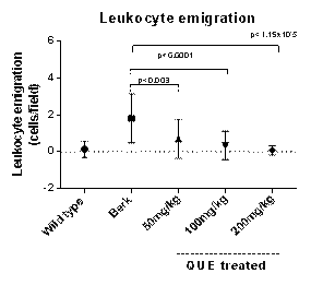
Results: QUE treatment restored blood flow, as evidenced by complete disappearance of vaso-occlusion in the postcapillary venules of Berk mice (Figure 1). However, no significant differences in venular diameter were noted with QUE treatment at any of the dose levels tested (50, 100, 200mg/kg) when compared to untreated Berk and wild type mice. But, when compared to untreated Berk mice, a significant increase in the RBC velocity was demonstrated in a dose dependent fashion (treated: 1.74 ±1.3 mm/sec, 3.02± 1.2 mm/sec, 3.4±0.90 mm/sec for 50, 100, 200 mg/kg dosing respectively vs. untreated 1.01± 1.05mm/sec, p<0.05). A dose of 200 mg level completely neutralized the vaso-occlusion. Increases in wall shear rate (650.01± 252.05 s-1 vs. 180.12± 165.02 s-1, p<6.03x10⁻⁶) was also observed in QUE treated vs. untreated Berk. This improvement of blood flow in the postcapillary venules correlated well with observed decreases in leukocyte adhesion (Figure 2A) and leukocyte emigration (Figure 2B) in QUE treated Berk mice (for doses 50, 100, and 200mg/kg) when compared to untreated Berk mice. Leukocyte rolling was also decreased for doses 100 and 200mg/kg (p<0.007, p<0.0002 respectively) after treatment with QUE when compared to untreated Berk and wild type.
Representative images showing postcapillary venules in the cremaster muscle microcirculation of Berk mice compared to QUE treated and wild type. Black arrows indicate leukocytes and white arrows indicate the blood flow direction.
Representative images showing postcapillary venules in the cremaster muscle microcirculation of Berk mice compared to QUE treated and wild type. Black arrows indicate leukocytes and white arrows indicate the blood flow direction.
Leukocyte adhesion (2A) and emigration (2B) in QUE treated Berk mice at 50, 100 and 200mg/kg doses compared to untreated Berk and wild type.
Leukocyte adhesion (2A) and emigration (2B) in QUE treated Berk mice at 50, 100 and 200mg/kg doses compared to untreated Berk and wild type.
Conclusion: We observed an improvement in RBC velocity and wall shear rate, as well as a complete attenuation of leukocyte adhesion, rolling and emigration at the highest dose of QUE treated transgenic sickle Berk mice. We suggest that these effects may be due to a decreased sickle RBC interaction with the vascular bed. Our present data provide a strong basis for the therapeutic application of flavonoids in SCD. Further studies are needed to better understand the mechanism of action in vivo for therapeutic effect in SCD.
Thangaswamy:AMI Life Sciences Private Ltd: Drug supplied Other. Billett:Selexys Pharmaceuticals: Research Funding.
Author notes
Asterisk with author names denotes non-ASH members.


This feature is available to Subscribers Only
Sign In or Create an Account Close Modal