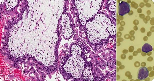
Thirteen months after the successful treatment of a persistent complete hydatidiform mole (panel A: diffusely hydropic, grape-like chorionic villi surrounded by hyperplastic atypical trophoblast), a 27-year-old woman with no family history of cancer presented with fatigue, sore throat, and petechiae for a few weeks. Her prior treatment included a single cycle of single-agent methotrexate that was followed, because of a further increase in human chorionic gonadotropin, by 9 biweekly cycles of etoposide, methotrexate, dactinomycin, cyclophosphamide, and vincristine (EMA/CO regimen). Blood work at this time showed hemoglobin, 10.9 g/dL; white blood cells, 41.4 × 10⁹/L; platelets, 15 × 109/L; and 60% blasts (panel B). The bone marrow was packed (95% cellularity) with immature myeloid cells that stained positively for CD13, CD33, CD34, and myeloperoxidase and negatively for CD3. Cytogenetic analysis revealed a core-binding factor β rearrangement at 16q22. A diagnosis of therapy-related acute myeloid leukemia (t-AML) was established.
Although t-AML after exposure to alkylating agents has a latency of 5 to 7 years, t-AML after exposure to topoisomerase II (TopoII) inhibitors has a shorter latency (1-3 years). Containing 2 TopoII inhibitors, 1 alkylating agent, 1 antitubilin agent, and 1 antimetabolite, EMA/CO carries a high risk of about 0.7% for future t-AML, and patients need close follow-up for several years after treatment.
For additional images, visit the ASH IMAGE BANK, a reference and teaching tool that is continually updated with new atlas and case study images. For more information visit http://imagebank.hematology.org.


This feature is available to Subscribers Only
Sign In or Create an Account Close Modal