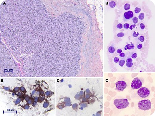
A 48-year-old woman without a familial history of multiple endocrine neoplasia presented with cervical pain. A palpable thyroid nodule was found surrounded by enlarged lymph nodes. A high serum level of calcitonin (560 pg/mL, normal < 8) and low calcium evoked the idea of a medullary thyroid carcinoma (MTC). Serum carcinoembryonic antigen (CEA) was 7.5 ng/mL (normal < 4). Histopathological examination demonstrated sheets of polygonal cells with pseudoglandular arrangement and angioinvasion (hematoxylin-eosin; panel A). Despite extensive surgery, the disease progressed and metastasized to the liver. One year later, unexpected pancytopenia led to an investigation of the bone marrow. The infiltration was total, composed of clumps of round to spindle cells separated by amorpheous deposits (May-Grünwald-Giemsa; panels B-C). Immunohistochemistry showed positive tumor cells for calcitonin, chromogranin A (panel D), and synaptophysin (panel E), confirming the involvement of parafollicular calcitonin-producing C-cells. Calcitonin and CEA levels were increased to 316 pg/mL and 1723 ng/mL, respectively. The patient was treated by chemotherapy but soon died.
MTC represents <10% of all primary thyroid malignancies and may occur sporadically or in the context of type 2 multiple endocrine neoplasia. Tumor cells derive from C-cells. If local metastatic extension to the cervical nodes is frequent, distant metastasis to the lung, liver, and bone are rare, and the growth usually remains slow. Extensive surgery is the standard treatment of local forms, but targeted therapy is a promising alternative for widespread disease.
For additional images, visit the ASH IMAGE BANK, a reference and teaching tool that is continually updated with new atlas and case study images. For more information visit http://imagebank.hematology.org.


This feature is available to Subscribers Only
Sign In or Create an Account Close Modal