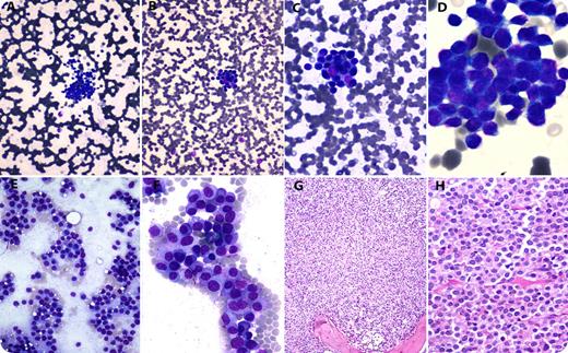
A 55-year-old man was admitted with fever and rapidly progressive renal failure with serum creatinine levels of 6.5 mg/dL and blood urea of 98 mg/dL. Complete blood counts revealed a hemoglobin of 50 g/L, a leukocyte count of 13.3 × 10⁹/L, and a platelet count of 46 × 109/L. Peripheral blood smear examination showed clusters of cells dispersed throughout the smear (panels A-D). These clusters were composed of mononuclear cells, with a rounded nucleus and a scant to moderate amount of basophilic cytoplasm. Occasional circulating myelocyte was also noted. A possibility of carcinocythemia was suggested, and in addition to investigations for the search of primary malignancy, a bone marrow examination was advised. The bone marrow aspirate smears (panels E-F) and trephine biopsy (panels G-H) showed replacement of normal hematopoietic cells by sheets of mature and immature plasma cells. Immunophenotyping on flow cytometry showed a surface expression of CD45 and CD38. The cells were negative for CD19, CD20, CD79b, FMC7, CD22, CD10, CD3, CD5, CD4, CD8, CD138, and CD56. Serum electrophoresis showed the presence of an M-band. Overall, the features were consistent with plasma cell leukemia. The patient succumbed to illness before any further workup or active treatment intervention.
Although rare, this case demonstrates that myeloma cells may cluster in the peripheral blood, similar to tumor cell clusters with carcinoma. The bone marrow biopsy correctly diagnosed myeloma as the cause of the clusters in this case.
For additional images, visit the ASH IMAGE BANK, a reference and teaching tool that is continually updated with new atlas and case study images. For more information visit http://imagebank.hematology.org.


This feature is available to Subscribers Only
Sign In or Create an Account Close Modal