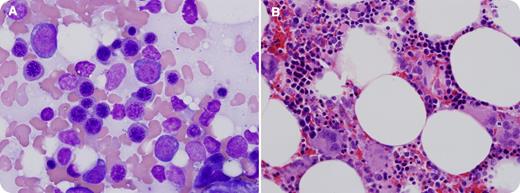
A 73-year-old white male presented to his primary care physician with abdominal pain, vomiting, dyspnea, and dysphagia lasting 1 week. He also reported white patches in his mouth and a cough that produced white sputum that started during this time period. A complete blood count with differential demonstrated severe neutropenia with a white blood cell count of 0.4 × 10⁹/L (absolute neutrophil count, 0 × 109/L; absolute lymphocyte count, 0.3 × 10⁹/L). The peripheral blood smear showed marked leukopenia with no blasts identified. Platelet count was 419 × 109/L and hemoglobin was 108 g/L. On physical examination, the patient was found to be in atrial fibrillation and oral candidiasis was noted. A chest radiograph showed a large mediastinal mass. Computed tomography of the chest showed a 6.6-cm × 4.4-cm necrotic anterior mediastinal mass that involved the phrenic nerve. Bone marrow biopsy revealed hypocellular bone marrow with granulocytic aplasia. Wright-Geimsa stained bone marrow aspirate smear (1000×) with preserved erythroid precursors, but absent granulopoiesis (panel A). The H&E stained bone marrow biopsy sections (400×) similarly demonstrates islands of erythroid development and appropriately distributed megakaryocytes, without maturing granulocytes (panel B). The mass was biopsied and thymic carcinoma (type C thymoma), subtype 1.2.1, epidermoid keratinizing (squamous cell) carcinoma was identified.
Pure white cell aplasia is a rare but recognized complication of thymoma and thymic carcinoma. Severe neutropenia of unknown etiology should prompt evaluation for possible mediastinal mass.
For additional images, visit the ASH IMAGE BANK, a reference and teaching tool that is continually updated with new atlas and case study images. For more information visit http://imagebank.hematology.org.


This feature is available to Subscribers Only
Sign In or Create an Account Close Modal