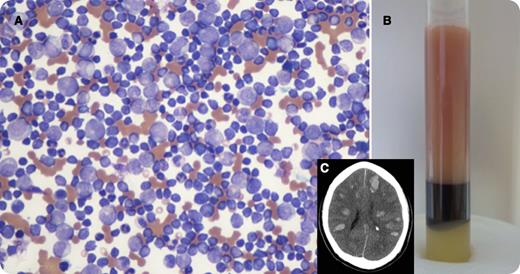
A 56-year-old male with no known past medical history presented with right-sided weakness, altered mental status, and acute respiratory failure. On presentation, his white blood cell count was 662 000/mm3, hemoglobin was 6.4 g/dL, and platelets were 20 000/mm3, with ∼80% blasts. Panel A is a blood smear obtained on presentation and panel B is the patient’s peripheral blood in a tube demonstrating his markedly elevated leukocrit relative to his hematocrit. Following intubation, emergent leukapheresis was performed, which resulted in a >50% reduction of his white blood cell count. His mental status continued to deteriorate with the absence of brain stem reflexes. A CT scan of the head was performed (panel C), which revealed numerous bilateral intracranial hemorrhages. The patient died shortly after admission secondary to massive cerebral bleed. Peripheral blood flow cytometry confirmed the diagnosis of an acute precursor B-lymphoblastic leukemia with >95% of cells positive for CD10, CD19, CD34, CD38, and HLA-DR. The interphase fluorescence in situ hybridization was positive for t(9;22)/BCR-ABL1 in >90% of the cells.
A 56-year-old male with no known past medical history presented with right-sided weakness, altered mental status, and acute respiratory failure. On presentation, his white blood cell count was 662 000/mm3, hemoglobin was 6.4 g/dL, and platelets were 20 000/mm3, with ∼80% blasts. Panel A is a blood smear obtained on presentation and panel B is the patient’s peripheral blood in a tube demonstrating his markedly elevated leukocrit relative to his hematocrit. Following intubation, emergent leukapheresis was performed, which resulted in a >50% reduction of his white blood cell count. His mental status continued to deteriorate with the absence of brain stem reflexes. A CT scan of the head was performed (panel C), which revealed numerous bilateral intracranial hemorrhages. The patient died shortly after admission secondary to massive cerebral bleed. Peripheral blood flow cytometry confirmed the diagnosis of an acute precursor B-lymphoblastic leukemia with >95% of cells positive for CD10, CD19, CD34, CD38, and HLA-DR. The interphase fluorescence in situ hybridization was positive for t(9;22)/BCR-ABL1 in >90% of the cells.
For additional images, visit the ASH IMAGE BANK, a reference and teaching tool that is continually updated with new atlas and case study images. For more information visit http://imagebank.hematology.org.


This feature is available to Subscribers Only
Sign In or Create an Account Close Modal