In this issue of Blood, Zhang and colleagues report the identification of a novel subset of circulating myeloid cells with immunosuppressive activity in pediatric patients with metastatic sarcomas.1
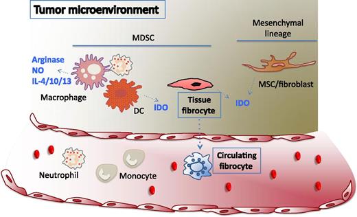
Tumor promotes the recruitment of myeloid- and mesenchymal-derived suppressor cells. Tumor formation is associated with the recruitment of cells of myeloid and mesenchymal origin that, as a consequence of the tumor secretome, are polarized toward an antiinflammatory and immunosuppressive profile. Circulating fibrocytes detected in patients with metastatic tumor display features of the myeloid compartment and exert immunosuppressive activity in vitro. Because of these characteristics, fibrocytes can be affiliated to the group of MDSCs. The immunosuppressive activity can be attributed to the production of IDO, which depletes the cellular microenvironment of tryptophan, required for T-cell proliferation. Despite substantial differences in marker expression and lineage of origin, IDO is also the main mechanism by which tumor-polarized DCs or MSCs inhibit immune responses. In contrast, polarized monocytes, macrophages, and neutrophils preferentially use a different set of molecules including arginase, nitric oxide, interleukin (IL)-4, IL-10, and IL-13.
Tumor promotes the recruitment of myeloid- and mesenchymal-derived suppressor cells. Tumor formation is associated with the recruitment of cells of myeloid and mesenchymal origin that, as a consequence of the tumor secretome, are polarized toward an antiinflammatory and immunosuppressive profile. Circulating fibrocytes detected in patients with metastatic tumor display features of the myeloid compartment and exert immunosuppressive activity in vitro. Because of these characteristics, fibrocytes can be affiliated to the group of MDSCs. The immunosuppressive activity can be attributed to the production of IDO, which depletes the cellular microenvironment of tryptophan, required for T-cell proliferation. Despite substantial differences in marker expression and lineage of origin, IDO is also the main mechanism by which tumor-polarized DCs or MSCs inhibit immune responses. In contrast, polarized monocytes, macrophages, and neutrophils preferentially use a different set of molecules including arginase, nitric oxide, interleukin (IL)-4, IL-10, and IL-13.
The last decade has witnessed a fundamental rethinking of the traditional concept of tumor-immune evasion. From the sole actor fighting host immune cells, tumor is now being seen as an entity deviously hijacking host immunoregulatory cells to create an immunoprivileged niche.
Different players have been identified among this class of cells that, depending on the need to be activated by an antigen receptor, can be classified into effectors of adaptive or innate tolerance. Regulatory T cells (Tregs) are probably the most studied in the first category, and their presence has been extensively documented in solid and hematologic malignancies, in which they are associated with a worse clinical outcome.2 More recently, B-cell regulatory subsets have also been observed in neoplastic conditions.
However, the most crowded field of investigation has been in the identification of innate immunity cell populations recruited by the tumor and instructed to acquire nonspecific immunosuppressive functions. They are cells of different lineage and tissue origin that can be classified into 2 major groups.
The first group consists of stromal cells of mesenchymal origin (mesenchymal stem/stromal cells [MSCs]), also considered progenitors of fibroblasts, or at least intimately related to them.3 MSCs/fibroblasts exhibit a potent immunosuppressive activity on virtually every cell type of the immune system4 and are the most abundant component of the solid tumor microenvironment. They actively contribute to malignant transformation,5 and their in vivo depletion restores antitumor immune responses.6
The second group comprises a heterogeneous group of myeloid cells that encompasses a large spectrum including macrophages, dendritic cells (DCs), and neutrophils.7 After appropriate activation, DCs are potent stimulators of immune responses, but when exposed to the tumor environment, they are converted to actively suppress T-cell functions. Closely related to DCs are macrophages, a major component of the leukocyte infiltrate of tumors. During tumor progression, the functional profile of tumor-associated macrophages is polarized from proinflammatory (M1) to antiinflammatory (M2) and is associated with the suppression of immune responses and the promotion of cancer growth. Similar to macrophages, neutrophils have been shown to shift from an antitumor (N1) phenotype to a protumor (N2) phenotype in the cancer environment in a transforming growth factor-β–dependent fashion.8 The overall group of myeloid cells exhibiting antiinflammatory/immunosuppressive qualities can be described as myeloid-derived suppressor cells (MDSCs).7
Despite major differences in their ontogeny, lineage, and differentiation stage, the players in both groups share fundamental similarities. First, all are functionally plastic because the microenvironment to which they are exposed determines their inflammatory profile. Second, the mechanisms underpinning their immunosuppressive activity often involve amino acid metabolic pathways and oxidative stress.7,9 Finally, as a downstream effect, most of these cellular pathways have the ability to activate Tregs.
Now Zhang et al report that fibrocytes can be a new player in the cancer-associated “innate tolerance” network. Fibrocytes, a cell subset of hematopoietic origin with fibroblast markers and morphology, had so far been considered inflammatory cells with a prominent role in the genesis of fibrosis.10 This study describes their potential role in effecting tumor evasion. The authors found increased numbers of circulating fibrocytes (CD14–CD11chiCD123–) in patients with metastasis. These fibrocytes expressed a phenotype resembling the one of MDSCs (CD11b+CD15+CD66b+IL4R+), but relatively distinct because of the high expression of HLA-DR and mesenchymal markers. Circulating fibrocytes from the patients, but not from normal individuals, exhibited immunosuppressive and proangiogenic activities, thus strongly suggesting the case for tumor-induced polarization and a further mechanism in tumor evasion from immune surveillance. Such a notion is consistent with the observation that fibrocyte differentiation decreases in inflammatory conditions and tissue injury, whereas it increases under conditions associated with wound healing and tissue remodeling.
Although not investigated in the study, it seems plausible that the nature of the “licensing” signals required for immunosuppressive fibrocytes (F2) are similar to those described for suppressors of myeloid or mesenchymal origin, such as tumor necrosis factor-α, interferon-γ, and the hypoxic tumor environment. The authors convincingly demonstrate that IL-4 can generate F2 in vitro, but the actual contribution of the tumor to their formation remains to be elucidated. It is also unknown whether fibrocytes at the tumor site exhibit the same characteristics of the circulating fibrocytes.6 A further similarity with the other suppressors is the mechanism of action. As with MSCs and tolerogenic DCs, fibrocytes use indoleamine 2-3 dioxygenase (IDO), which depletes the cellular microenvironment of the essential amino acid tryptophan, required for T-cell proliferation. The authors failed to detect any of the pathways involved in MDSC-mediated immune inhibition (arginase or nitric oxide), but this may depend on the cancer type, which may activate selective pathways only. Further analysis of the immunosuppressive molecules will unravel additional, potentially overlapping, mechanisms such as inhibitory cytokines (IL-4, IL-10, IL-13) and the ability to induce Tregs.
A question stemming from this study is whether fibrocytes, MSCs, and MDSCs are unique entities or whether they instead reflect a functional status—elicited by inflammation or tumor environment—that can be acquired by cells of different origin that have in common the machinery to activate innate pathways of immunosuppression. What we have learned from the characterization of the MDSC subsets suggests that tumor conditioning is more complex than a simple polarizing process, because each subset responds differently, and the same effector molecule can mediate different activities when it is secreted by different subsets.
From a translational perspective, the findings by Zhang and colleagues might add discouragement in regard to tumor immunotherapies because of the apparent need to control a new population. However, the evidence that tumor-associated fibrocytes use a well-known weapon (IDO) in the armamentarium of innate tolerance suggests that tumor can teach a new dog, but only old tricks.
Conflict-of-interest disclosure: The author declares no competing financial interests.


This feature is available to Subscribers Only
Sign In or Create an Account Close Modal