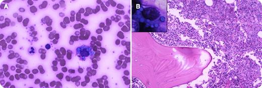
A 58-year-old white female had severe bone pain for 1 week. Increased levels of free λ light chain (2423 mg/L) were present in the serum with decreased levels of all immunoglobulins. The complete blood count and differential were within normal limits. The peripheral blood smear had increased rouleaux formation and atypical plasma cells containing numerous cytoplasmic Russell bodies (panel A). There were no lytic lesions on skeletal survey or positron emission tomography–computed tomography scan, and organmegaly or lymphadenopathy were not detected. Bone marrow biopsy was hypercellular with an extensive infiltrate of atypical plasma cells containing numerous small intracytoplasmic globules (Russell bodies); larger extracellular globules were also present (panel B). The marrow smear showed multiple blue intracytoplasmic Russell bodies, resembling a bunch of grapes, referred to as Mott or morula cells (panel B inset).
Flow cytometry and immunohistochemistry confirmed the λ light chain restriction in plasma cells. In addition, there was osteosclerosis in the bony trabeculae. A remarkable feature of the present case was the abundance of Russell bodies in the marrow as well as circulating plasma cells in a patient with λ light chain osteosclerotic myeloma.
For additional images, visit the ASH IMAGE BANK, a reference and teaching tool that is continually updated with new atlas and case study images. For more information visit http://imagebank.hematology.org.


This feature is available to Subscribers Only
Sign In or Create an Account Close Modal