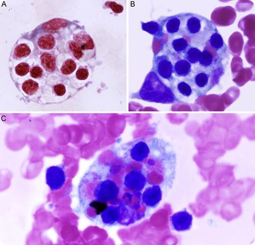
A 73-year-old male with a medical history of chronic lymphocytic leukemia (CLL) with urinary infection presented with fever (101.6°F) and pancytopenia (white blood cells 1.0 K/μL, platelets 54 K/μL, and hemoglobin 7.8 g/dL). Viral and fungal studies were negative. A bone marrow study was performed and showed persistent CLL and the presence of numerous phagocytosed lymphocytes and red blood cells by histiocytes. The figure shows characteristic engulfed red blood cells and lymphocytes by histiocytes (panel A: iron stain; panels B-C: Wright-Giemsa stain). Further laboratory tests demonstrated elevated ferritin (45 077 mg/dL), hypertriglyceridemia (4 mmol/L), hypophosphatemia (0.5 g/L), and hepatocellular pattern of transaminitis. The clinicopathological findings of this patient met the diagnostic criteria of hemophagocytic syndrome or hemophagocytic lymphohistiocytosis (HLH), a systemic, progressive, and often fatal disorder. Clinical improvement was observed after the patient was treated with etoposide and dexamethasone.
Two major forms of HLH have been described, familial (FHLH) and secondary or acquired (AHLH). FHLH is an autosomal recessive disorder prevalently seen in parental consanguinity. AHLH occurs secondary to systemic infection, immunodeficiency, and underlying malignancy, such as CLL in this patient. Treatment with immunosuppressive and/or chemotherapeutic agents might alleviate the disorder, although poor clinical outcomes of HLH were reported when associated with hematologic malignancies.
For additional images, visit the ASH IMAGE BANK, a reference and teaching tool that is continually updated with new atlas and case study images. For more information visit http://imagebank.hematology.org.


This feature is available to Subscribers Only
Sign In or Create an Account Close Modal