Key Points
First report demonstrating in vivo elimination of multiple LIC populations from childhood ALL cases using animal models.
In vivo models of leukemia are essential for drug evaluation studies.
Abstract
Approximately 20% of children with acute lymphoblastic leukemia (ALL) relapse because of failure to eradicate the disease. Current drug efficacy studies focus on reducing leukemia cell burden. However, if drugs have limited effects on leukemia-initiating cells (LICs), then these cells may expand and eventually cause relapse. Parthenolide (PTL) has been shown to cause apoptosis of LIC in acute myeloid leukemia. In the present study, we assessed the effects of PTL on LIC populations in childhood ALL. Apoptosis assays demonstrated that PTL was effective against bulk B- and T-ALL cells, whereas the CD34+/CD19−, CD34+/CD7−, and CD34− subpopulations were more resistant. However, functional analyses revealed that PTL treatment prevented engraftment of multiple LIC populations in NOD/LtSz-scid IL-2Rγc–null mice. PTL treatment of mice with established leukemias from low- and high-risk patients resulted in survival and restoration of normal murine hemopoiesis. In only 3 cases, disease progression was significantly slowed in mice engrafted with CD34+/CD19− or CD34+/CD7− and CD34− cells, but was not prevented, demonstrating that individual LIC populations within patients have different responses to therapy. These observations indicate that PTL may have therapeutic potential in childhood ALL and provide a basis for developing effective therapies that eradicate all LIC populations to prevent disease progression and reduce relapse.
Introduction
Childhood acute lymphoblastic leukemia (ALL) is a heterogeneous disease in terms of karyotype, immunophenotype, and blast morphology.1-3 Several genome-wide analyses have described additional genetic changes within individual leukemia clones.4-6 Therefore, studies that increase our understanding of the biology and evolution of this disease should provide information on leukemogenic pathways for therapeutic targeting. Such studies may help us to understand why initial therapies can induce remission but some patients then relapse,7-9 especially because many relapses occur in the low-risk groups.
ALL cells that can generate leukemias in immune-deficient mice, termed leukemia-initiating cells (LICs), were initially thought to be rare and have a hierarchical structure.10-13 Some of these LICs, particularly CD133+/CD19− cells, were found to be resistant to treatment with dexamethasone and vincristine,13 which are commonly used in the induction phase of therapy for ALL, so they may survive therapy and eventually cause disease relapse. However, more recent studies using NOD/LtSz-scid IL-2Rγc–null (NSG) mice have revealed that both primitive and more differentiated ALL subpopulations can initiate and maintain acute leukemias in this strain.5,14-18 We and others have shown that CD34+/CD19+, CD34+/CD19−, and CD34− B-cell precursor ALL subpopulations contain LICs.15,18 In childhood T-ALL, we found that the CD34+/CD7+, CD34+/CD7−, and CD34− subpopulations had LIC properties.18 Another study described CD7+/CD1a− and CD7+/CD1a+ LIC from 1 pediatric and 2 adult T-ALL patients,19 and a study of mature/cortical T-ALL found LICs in both the CD34+/CD7+ and CD34−/CD7+ populations.20 The in vivo repopulating capacity has been shown to vary depending on the immunophenotype18 and genotype of the LIC.5 These findings demonstrate that LICs are more abundant than earlier studies suggested and indicate that leukemia evolution may have a branching structure rather than a hierarchical one. This has significant implications for therapy of leukemias. Elimination of the population that contains the greatest proportion of LICs or has the greatest self-renewal potential may just lead to another LIC population evolving and expanding to maintain the disease. Therefore, it will be important to develop therapies that can eliminate all populations with LIC potential to prevent further evolution and recurrence. The challenge is to find a way to specifically target LICs without causing toxicity to normal cells.
Parthenolide (PTL) has been investigated recently as a potential chemotherapeutic agent for acute myeloid leukemia (AML) and chronic lymphocytic leukemia.21-26 PTL is a naturally occurring sesquiterpene lactone used in the treatment of fever, migraines, and rheumatoid arthritis and as an anti-inflammatory agent.27-29 PTL can induce apoptosis through a range of responses, such as inhibition of NF-κB, p53 activation, and increase of reactive oxygen species.21,23 PTL and DMAPT, an analog of PTL, have been shown to be effective in AML while sparing normal hemopoietic stem cells (HSCs).21,22 PTL can induce apoptosis in primary B-ALL cells22 and cell lines.30,31 To date, there are no reports on the effects of PTL on LIC populations in pediatric ALL. The aim of the present study was to evaluate the effects of PTL on childhood ALL cells, especially subpopulations that have LIC activity and including those that have been shown to be resistant to current therapeutic agents.13
Methods
Patient cells
BM cells from children (median age, 6 years and 8 months; range, 6 months to 19 years) with B-cell precursor ALL and T-ALL at presentation or relapse were collected in accordance with the Declaration of Helsinki and with approval of University Hospitals Bristol National Health Service Trust. Detailed characteristics of the patient samples are shown in Table 1. Samples were selected on the basis of availability of material for study only. Normal BM and peripheral blood (PB) samples were obtained from consenting healthy donors. Cells were separated using Ficoll-Hypaque (Sigma-Aldrich). Mononuclear cells were suspended in IMDM (Invitrogen) with 90% FCS (Invitrogen) and 10% DMSO (Manor Park Pharmaceuticals) and stored in liquid nitrogen. Mean viability of samples on thawing was 83% ± 17% for ALL samples and 71% ± 17% for normal samples.
Patient characteristics
| Patient no. . | Subtype . | Karyotype . | Age at diagnosis, y . | Sex . | Disease status at biopsy . | MRD risk status† . |
|---|---|---|---|---|---|---|
| 1 | c-ALL | 46XX | 5 | F | Diagnosis | ND |
| 2 | c-ALL | 46XY | 6 | M | Diagnosis | ND |
| 3 | Pre-B | t(4;11), +X | 0.6 | M | Diagnosis | ND |
| 4 | Pre-B | -9 | 7 | F | Diagnosis | High |
| 5 | c-ALL | t(12;21), −3, −6, +10 | 5 | F | Diagnosis | Low |
| 6 | Pro-B | t(4;11) | 0.5 | F | Relapse | High |
| 7 | Pre-B* | +21, +22 | 3 | M | Diagnosis | Low |
| 8 | c-ALL | -12p | 15 | M | Diagnosis | High |
| 9 | c-ALL | t(12;21), −12p | 6 | M | Diagnosis | IM |
| 10 | c-ALL | t(12;21) | 6 | M | Diagnosis | Low |
| 11 | c-ALL | t(8;21), +9 | 19 | F | Relapse | High |
| 12 | Pre-B | Complex | 14 | M | Diagnosis | Low |
| 13 | c-ALL | 46XX | 4 | F | Diagnosis | Low |
| 14 | Pro-B | t(12;17) | 14 | F | Diagnosis | High |
| 15 | c-ALL | t(12;21) | 4 | F | Diagnosis | Low |
| 16 | c-ALL | Hyperdiploid | 3 | F | Diagnosis | Low |
| 17 | Pro-B | t(4;11) | 0.8 | F | Diagnosis | High |
| 18 | c-ALL | i(9), del(9) | 16 | M | Relapse | Low |
| 19 | c-ALL | +7, +9, −12, (iAMP21) | 8 | F | Diagnosis | High |
| 20 | c-ALL | +3 | 14 | F | Diagnosis | High |
| 21 | Pre-B | t(12;21) | 8 | F | Diagnosis | High |
| 22 | Pre-B | Hyperdiploid | 3 | M | Relapse | Low |
| 23 | Pre-B | del 1 | 2 | F | Diagnosis | Low |
| 24 | Pre-B | Hyperdiploid | 2 | M | Diagnosis | Low |
| 25 | Pre-B | +14 | 2 | F | Diagnosis | Low |
| 26 | Pre-B | t(1;19) | 14 | M | Diagnosis | Low |
| 27 | Pre-B | t(9;22) | 15 | M | Diagnosis | Low |
| 28 | Pre-B | 46XY | 9 | M | Diagnosis | High |
| 29 | T-ALL | +4, +9 | 15 | M | Diagnosis | High |
| 30 | T-ALL | t(11;14) | 2 | M | Relapse | Low |
| 31 | T-ALL | -9p, +9q | 6 | M | Diagnosis | High |
| 32 | T-ALL | +7, −9, t(9;16) | 5 | M | Diagnosis | High |
| 33 | T-ALL | t(1;14), −6 | 10 | M | Diagnosis | Low |
| 34 | T-ALL | 46XY | 14 | M | Diagnosis | High |
| 35 | T-ALL | t(6;7) | 1 | M | Diagnosis | High |
| 36 | T-ALL | -6 | 15 | M | Diagnosis | High |
| 37 | T-ALL | 46XY | 13 | M | Diagnosis | High |
IM indicates intermediate; ND, not determined; and MRD, minimal residual disease.
ALL secondary to Down syndrome.
MRD risk status at day 28.
Cell sorting
B-ALL cells were stained with antibodies against CD34 (clone 8G12) and CD19 (clone 4G7). T-ALL cells were stained with anti-CD34 and anti-CD7 (clone M-T701). Normal BM and PB samples were stained with anti-CD34 and anti-CD38 (clone HB7). IgG1 antibodies were used as isotype controls (all from BD Biosciences). Cells were sorted using an Influx cell sorter (BD Biosciences) with Spigot Version 6.1.9 software on the basis of fluorescence intensity after gating on 7-aminoactinomycin D (Sigma-Aldrich)–negative cells with low forward and side scatter. Details of the proportions of nucleated cells in the sorted populations are provided in supplemental Table 1 (available on the Blood Web site; see the Supplemental Materials link at the top of the online article). Sorting was performed using the maximum purity setting and the purity of sorted subfractions from each sample was checked during and after sorting.
In vivo studies
Animal experiments were conducted under licenses approved by the United Kingdom Home Office. NSG mice were bred and maintained at the University of Bristol Animal Service Unit. Mice were not preconditioned before inoculation. Cells were resuspended in 0.3 mL of IMDM + 5% human albumin solution and injected into the lateral tail veins of 6- to 8-week-old mice. Unsorted cells and sorted cell populations were inoculated at a range of doses to obtain estimates of LIC frequency in each subpopulation.
In ex vivo drug sensitivity assays, unsorted cells and sorted subpopulations from ALL patients and healthy donors were treated with either PTL for 20-24 hours or DMSO + PBS (Invitrogen) as controls before inoculation.
In survival assays, mice were inoculated with bulk ALL cells and sorted subpopulations. Mice were monitored weekly for the presence of human cells in PB aspirates. Once the level of human cells was ≥ 5%, mice were given daily IV doses of PTL (40 mg/kg) or placebo (DMSO + PBS) for 9 days.22,32
In all assays, mice were monitored weekly for the presence of human cells, maintained for up to 20 weeks, and killed electively or when they began to exhibit clinical symptoms of disease. The gross anatomy was inspected and femoral BM samples were removed for flow cytometric and cytogenetic/histologic analyses. Immunophenotypes of xenografts were examined using antibodies against human CD3, CD10, CD19, CD7, CD33, CD34, CD45, and murine CD45 (all BD Biosciences). Cytogenetic analysis by FISH was performed by Bristol Genetics Laboratory, Southmead Hospital, Bristol.
Cells harvested from the BM of some engrafted mice were used for serial transplantation experiments. For comparison with primary transplantations, equal numbers of human CD45 cells were inoculated into serial recipients. These cells were not enriched for any particular phenotype before evaluation in sequential xenografts.
In vitro drug sensitivity
Unsorted ALL cells and sorted subpopulations were cocultured with increasing concentrations of PTL (Sigma-Aldrich) for 20-24 hours in RPMI 1640 medium (Sigma-Aldrich) containing 10% FCS and 1% l-glutamine (Invitrogen). Unsorted and HSC-enriched (CD34+/CD38−) normal BM samples were also cocultured with the drug. Apoptosis and viability were assessed by flow cytometry using annexin V–FITC and propidium iodide (arcus biologicals).
Western blotting and confocal microscopy
Cells for Western blotting were prepared using the method described by Foka et al.33 Full details are provided in supplemental Methods.
Statistical analysis
LIC frequencies were determined by Poisson statistics using L-Calc Version 1.1 software (StemCell Technologies). ANOVA, followed by Tukey posthoc testing, was used to compare drug responses between ≥ 3 populations. Matched paired t tests were used to compare viability and engraftment levels between untreated and drug-treated groups. The t tests assuming unequal variance were used to compare drug-treated ALL populations with treated HSCs. Data from survival assays were analyzed using the log-rank test of survival distribution after treatment.
Results
LIC frequency in ALL subpopulations
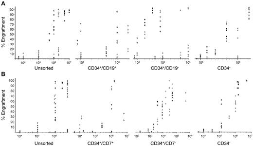
Cells from 15 cases were sorted based on expression of CD34 and CD19 (B-ALL cases, Figure 1A) and CD34 and CD7 (T-ALL cases, Figure 1B). Cells were inoculated at a range of doses to give an estimate of the frequency of LICs (Table 2). LIC frequency in sorted subpopulations varied from 1 in 700 to 1 in 9 × 10⁵, but was highest in CD34+/CD19− cells in B-ALL cases and CD34+/CD7− cells in T-ALL cases. Because the number of sorted cells available limited the extent of the dilution analysis, these results are likely to be an underestimate of LIC frequencies. Because LICs were readily detectable in all sorted subfractions, we subsequently assessed the effects of PTL on these subpopulations.
Assessment of the LIC content of sorted ALL subpopulations. B-ALL cells from 8 patients (patients 2, 6, 16, 17, 20, 22, 23, and 28) were sorted for expression of CD34 and CD19 (A) and cells from 7 T-ALL patients (patients 29-32 and 34-36) were sorted for expression of CD34 and CD7 (B). Unsorted cells and sorted subpopulations were inoculated into NSG mice to evaluate the LIC content. Two to 4 mice were injected per cell dose. Each patient is represented by a specific symbol and each symbol depicts the leukemia engraftment detected in the BM of an individual mouse.
Assessment of the LIC content of sorted ALL subpopulations. B-ALL cells from 8 patients (patients 2, 6, 16, 17, 20, 22, 23, and 28) were sorted for expression of CD34 and CD19 (A) and cells from 7 T-ALL patients (patients 29-32 and 34-36) were sorted for expression of CD34 and CD7 (B). Unsorted cells and sorted subpopulations were inoculated into NSG mice to evaluate the LIC content. Two to 4 mice were injected per cell dose. Each patient is represented by a specific symbol and each symbol depicts the leukemia engraftment detected in the BM of an individual mouse.
Frequency of LICs in ALL subpopulations
| Patient no. . | Subpopulation . | |||
|---|---|---|---|---|
| Unsorted . | CD34+/CD19+ . | CD34+/CD19− . | CD34 . | |
| 2 | 1 in 4 × 10⁴ | 1 in 2 × 10⁵ | 1 in 3 × 10³ | 1 in 2 × 10⁵ |
| 6 | < 1 in 1 × 10⁶ | 1 in 4 × 10⁵ | < 1 in 2 × 10³ | 1 in 1 × 10⁴ |
| 16 | 1 in 2 × 10⁵ | 1 in 2 × 10⁵ | 1 in 8 × 10³ | 1 in 3 × 10⁵ |
| 17 | 1 in 9 × 10⁴ | 1 in 4 × 10⁵ | 1 in 8 × 10² | 1 in 5 × 10⁵ |
| 20 | 1 in 5 × 10⁴ | 1 in 9 × 10⁴ | < 1 in 1 × 10³ | 1 in 3 × 10⁵ |
| 22 | 1 in 2 × 10⁴ | 1 in 7 × 10⁴ | 1 in 9 × 10² | 1 in 9 × 10⁴ |
| 23 | 1 in 7 × 10⁵ | 0 | 1 in 9 × 10³ | 0 |
| 28 | 1 in 7 × 10³ | < 1 in 8 × 10⁴ | 1 in 7 × 10² | 1 in 1 × 10⁵ |
| Cell dose | 5 × 10³ to 1 × 10⁷ | 8 × 10⁴ to 2 × 10⁶ | 8 × 10² to 1 × 10⁵ | 8 × 10⁴ to 2 × 10⁶ |
| Patient no. . | Subpopulation . | |||
|---|---|---|---|---|
| Unsorted . | CD34+/CD7+ . | CD34+/CD7− . | CD34− . | |
| 29 | 1 in 2 × 10⁵ | 1 in 2 × 10⁴ | 1 in 2 × 10³ | 1 in 3 × 10⁴ |
| 30 | 1 in 9 × 10³ | 1 in 6 × 10³ | 1 in 2 × 10³ | 1 in 2 × 10⁴ |
| 31 | 1 in 6 × 10⁵ | 1 in 8 × 10³ | 1 in 3 × 10³ | |
| 32 | 1 in 2 × 10⁵ | 1 in 9 × 10³ | 1 in 7 × 10² | 1 in 5 × 10⁵ |
| 34 | 1 in 2 × 10⁴ | 1 in 9 × 10⁴ | < 1 in 9 × 10² | 1 in 3 × 10⁴ |
| 35 | 1 in 9 × 10⁴ | 1 in 6 × 10⁴ | 1 in 1 × 10³ | 1 in 1 × 10⁴ |
| 36 | 1 in 1 × 10⁵ | 1 in 9 × 10⁵ | 1 in 5 × 10³ | 1 in 9 × 10⁴ |
| Cell dose | 5 × 10³ to 5 × 10⁶ | 4 × 10³ to 5 × 10⁶ | 2 × 10² to 5 × 10⁵ | 5 × 10³ to 5 × 10⁶ |
Viability of PTL-treated ALL cells
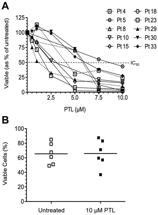
PTL was initially tested in vitro at a dose range from 0.5-10μM (Figure 2A) in 10 cases. The half-maximal inhibitory concentration (IC50) was reached at 7.5μM in 9 cases, but 10μM PTL reduced the viability of all cases to ≤ 43.3%. This higher dose had no effect on the viability of normal HSCs (P = .91, Figure 2B). Therefore, 10μM PTL was used routinely thereafter to maximize effects in all patients.
Response of ALL cells and normal HSCs to PTL. (A) Percentage of viable ALL cells after PTL treatment in 7 B-ALL patients (open symbols) and 3 T-ALL patients (closed symbols). PTL was used at a dose ranging from 0.5-10μM and viability was assessed at 20-24 hours by flow cytometry using annexin V and propidium iodide. Samples depicted with dashed lines are high minimal residual disease risk patients. (B) Viability of CD34+/CD38− HSCs from normal BM treated with PTL (10μM) compared with untreated controls (n = 6).
Response of ALL cells and normal HSCs to PTL. (A) Percentage of viable ALL cells after PTL treatment in 7 B-ALL patients (open symbols) and 3 T-ALL patients (closed symbols). PTL was used at a dose ranging from 0.5-10μM and viability was assessed at 20-24 hours by flow cytometry using annexin V and propidium iodide. Samples depicted with dashed lines are high minimal residual disease risk patients. (B) Viability of CD34+/CD38− HSCs from normal BM treated with PTL (10μM) compared with untreated controls (n = 6).
Unsorted cells from 16 of 23 B-ALL patients responded to the drug (< 38% viable), 4 showed a partial response (58%-71%) and 3 were largely unaffected (> 87% viable). There was a significant reduction in viability in all populations after treatment compared with untreated controls (P ≤ .043). When individual subpopulations were compared, CD34+/CD19− cells were the least affected, with 81.3% ± 36% surviving (P = .0005, F = 6.64, Fcrit = 2.71). Less than 42% ± 40% cells from unsorted samples or other subpopulations remained viable after treatment. Individual results are shown in supplemental Table 2.
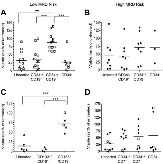
When B-ALL cases were grouped according to minimal residual disease risk status, where available, into low (n = 12) and high (n = 8) risk, a similar response pattern was observed in the low-risk group (Figure 3A), with CD34+/CD19− cells being the most resistant (87.6% ± 26% viable, P = .0001, F = 11.31, Fcrit = 2.82). The viability of the other subpopulations was ≤ 35% ± 33%, significantly lower than in HSCs (100% ± 14% viable, P ≤ .0004). In the high-risk group, there was no significant difference in viability between the sorted subpopulations (P = .53, F = 0.75, Fcrit = 2.90). High-risk cases were less responsive and CD34+/CD19− and CD34− subpopulations were the most resistant (71.3% ± 45% and 70.3% ± 51% viable, respectively). These responses were not significantly different from results obtained with HSCs (0.12 < P < .43). However, the viability of treated unsorted and CD34+/CD19+ cells was significantly lower than treated HSCs (P ≤ .013).
Viability of B- and T-ALL populations after PTL exposure. (A) Low minimal residual disease risk (MRD; n = 12) and (B) high MRD risk (n = 8) B-ALL subpopulations were sorted using antibodies against CD34 and CD19. (C) Six patients were sorted using antibodies against CD133 and CD19. Squares represent the low-risk patients; triangles, intermediate risk; and circles, high-risk. (D) Subpopulations from 9 T-ALL patients were sorted using antibodies against CD34 and CD7. Two of the 9 patients were low MRD risk and are depicted with open squares. High MRD risk patients are depicted with filled circles. Graphs show the proportion of viable B- and T-ALL subpopulations after exposure to 10μM PTL. Data are expressed as a percentage of untreated controls. Each symbol represents results from individual patients. Horizontal bars represent mean viability. **P ≤ .01; ***P ≤ .001.
Viability of B- and T-ALL populations after PTL exposure. (A) Low minimal residual disease risk (MRD; n = 12) and (B) high MRD risk (n = 8) B-ALL subpopulations were sorted using antibodies against CD34 and CD19. (C) Six patients were sorted using antibodies against CD133 and CD19. Squares represent the low-risk patients; triangles, intermediate risk; and circles, high-risk. (D) Subpopulations from 9 T-ALL patients were sorted using antibodies against CD34 and CD7. Two of the 9 patients were low MRD risk and are depicted with open squares. High MRD risk patients are depicted with filled circles. Graphs show the proportion of viable B- and T-ALL subpopulations after exposure to 10μM PTL. Data are expressed as a percentage of untreated controls. Each symbol represents results from individual patients. Horizontal bars represent mean viability. **P ≤ .01; ***P ≤ .001.
The effect of PTL was also investigated in B-ALL subpopulations using CD133 (Figure 3C and supplemental Table 3). CD133+/CD19+ and CD133+/CD19− subpopulations from 2 of the patients used in the previous paragraph (patients 8 and 15) and 4 additional patients (patients 4, 5, 9, and 18) were sorted and treated with 10μM PTL. In this cohort, a greater response to PTL was observed using unsorted cells (13.7% ± 17% viable) compared with the larger cohort used above, and only 5.9% ± 5% CD133+/CD19+ cells survived. PTL had a reduced effect on the CD133+/CD19− subpopulation (71.6% ± 21% viable) compared with the other populations assayed (P = .0002, F = 20.91, Fcrit = 4.10), but this was significantly reduced compared with results from HSCs (P = .032).
Seven of 9 T-ALL patients were affected by PTL (< 36% viable) and a partial response was observed in the 2 remaining cases (62%-69% viable, Figure 3D). Only 27.2% ± 27% of unsorted T-ALL cells survived treatment, whereas 48.4% ± 29% of CD34+/CD7+ cells, 54.9% ± 41% of CD34+/CD7− cells, and 57.8% ± 74% of CD34− cells remained viable (P = .53, F = 0.76, Fcrit = 3.01). When treated populations were compared with their untreated counterparts, there was a significant reduction in the viability in all populations (P ≤ .039), except in CD34− cells (P = .105). This less marked effect of PTL on CD34− cells was largely attributable to 2 patients in which these cells were unaffected by PTL, whereas in the remaining patients, < 16% of CD34− cells survived (P = .04 cf untreated). The proportions of unsorted CD34+/CD7+ and CD34+/CD7− T-ALL cells surviving PTL treatment were significantly reduced compared with HSCs (P ≤ .018).
In vivo engrafting capacity of PTL-treated cells
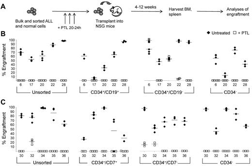
Unsorted cells and sorted subpopulations from 5 B-ALL patients and 5 T-ALL patients were treated with 10μM PTL for 20-24 hours before inoculation to assess whether PTL had any effects on the ability of cells to engraft NSG mice (Figure 4A). Groups of 2-4 mice were inoculated with each population assayed. Cell doses that had previously resulted in good engraftment levels were used. The number of sorted cells inoculated varied because of phenotypically primitive cells representing only small proportions of the total leukemia blast population (supplemental Table 1) and different immunophenotypes among patients. Treatment of unsorted CD34+/CD19+ and CD34− B-ALL cells with PTL before inoculation completely prevented engraftment in every case (Figure 4B). PTL treatment of CD34+/CD19− cells significantly reduced or prevented engraftment in 3 patients (patients 17, 22, and 28). In the other 2 patients, the engrafting capacity of CD34+/CD19− cells from patient 20 was unaffected and engraftment was higher in mice inoculated with PTL-treated cells from patient 6 (98% ± 3%) than with untreated cells (76% ± 12%, supplemental Figure 1). FISH analyses on cells recovered from mice engrafted with unsorted and all sorted subpopulations confirmed these cells had the same karyotype as the patients at diagnosis (91%-100% FISH+). Likewise, cells recovered from mice engrafted with PTL-treated CD34+/CD19− cells from patients 6 and 20 carried the patient-specific aberrations (95%-100% FISH+).
In vivo propagating ability of PTL-treated ALL subpopulations. (A) Unsorted and sorted ALL cells were treated with PTL for 20-24 hours in vitro, and then injected into NSG mice. Cells were inoculated at the following doses per mouse: unsorted (1-5 × 10⁶), CD34+/CD19+ (8 × 10⁴-1 × 10⁶), CD34+/CD19− (1.8 × 10³-1.2 × 10⁵), CD34− (1.5-2 × 10⁶ B-ALL), CD34+/CD7+ (1-1.5 × 10⁶), CD34+/CD7− (4-6 × 10⁴), and CD34− (1.5-5 × 10⁶ T-ALL). After 4-12 weeks, leukemia cell engraftment in BM was analyzed by flow cytometry using a panel of antibodies. (B-C) Levels of engraftment attained using unsorted and sorted subpopulations from 5 B-ALL patients (B) and from 5 T-ALL patients (C). Each symbol depicts the engraftment level of human leukemia cells from the patients indicated as measured in the BM of individual mice. Closed symbols represent mice inoculated with untreated cells; opens symbols, mice inoculated with PTL-treated cells. Solid horizontal bars represent mean engraftment levels using untreated cells; dashed bars represent mean engraftment levels using PTL-treated cells.
In vivo propagating ability of PTL-treated ALL subpopulations. (A) Unsorted and sorted ALL cells were treated with PTL for 20-24 hours in vitro, and then injected into NSG mice. Cells were inoculated at the following doses per mouse: unsorted (1-5 × 10⁶), CD34+/CD19+ (8 × 10⁴-1 × 10⁶), CD34+/CD19− (1.8 × 10³-1.2 × 10⁵), CD34− (1.5-2 × 10⁶ B-ALL), CD34+/CD7+ (1-1.5 × 10⁶), CD34+/CD7− (4-6 × 10⁴), and CD34− (1.5-5 × 10⁶ T-ALL). After 4-12 weeks, leukemia cell engraftment in BM was analyzed by flow cytometry using a panel of antibodies. (B-C) Levels of engraftment attained using unsorted and sorted subpopulations from 5 B-ALL patients (B) and from 5 T-ALL patients (C). Each symbol depicts the engraftment level of human leukemia cells from the patients indicated as measured in the BM of individual mice. Closed symbols represent mice inoculated with untreated cells; opens symbols, mice inoculated with PTL-treated cells. Solid horizontal bars represent mean engraftment levels using untreated cells; dashed bars represent mean engraftment levels using PTL-treated cells.
Treatment with PTL completely prevented engraftment of unsorted T-ALL cells in 4 of 5 patients (Figure 4C). In the remaining case (patient 32) engraftment was significantly reduced to 22% ± 6% compared with untreated cells (77% ± 9%, P < .001). No engraftment was observed using PTL-treated CD34+/CD7+ and CD34− cells. In 3 patients, PTL prevented engraftment of CD34+/CD7− cells and significantly reduced engraftment in 2 patients, from 98% ± 3% to 15% ± 4% (patient 30) and from 52% ± 7% to 16% ± 4% (patient 32), P < .001. FISH analyses confirmed that the engrafted cells had an aberrant karyotype (67%-100% FISH+). PTL treatment of normal CD34+/CD38− HSC before inoculation did not have any detrimental effects on the functional capacity of these cells. Engraftment levels of treated cells were 120% relative to the untreated controls (P = .25, supplemental Figure 2).
Self-renewal capacity of resistant cells
In the 3 patients in which in vitro PTL treatment of LICs had little (CD34+/CD19−, patients 6 and 20) or limited (CD34+/CD7−, patient 32) effects on engrafting capacity, cells recovered from murine BM were transplanted into secondary mice to assess the self-renewal ability of the PTL-treated cells (supplemental Table 4). Cells were not resorted before inoculation into secondary recipients, but they received equivalent numbers of human cells as their primary counterparts. Comparable levels of engraftment were observed in the primary and secondary recipients. Inoculated LICs differentiated in vivo to give rise to leukemias that were typical of the patient samples at biopsy. However, a proportion of the grafts from mice inoculated with PTL-treated or untreated CD34+/CD19− cells remained CD34+/CD19− (3.7% ± 0.6%). Likewise, the majority of cells recovered from mice inoculated with treated or untreated CD34+/CD7− cells were CD34+/CD7+, but 14.2% ± 2.3% remained CD34+/CD7−, indicating self-renewal of these primitive LICs.
In vivo activity of PTL in NSG mice
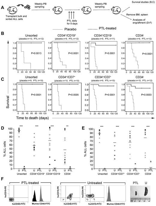
Unsorted and sorted cell populations from 9 patients were inoculated into NSG mice and PTL was administered when leukemia was established (Figure 5A). Disease progression was prevented in PTL-treated mice, whereas the leukemia burden in placebo-treated mice continued to increase until they had all succumbed to disease within 9 weeks after treatment commenced. PTL significantly increased survival for each treated xenograft (P < .0014, Figure 5B-C). The median survival time of untreated mice was 29 days (range, 2-60) after treatment commenced for those engrafted with sorted and unsorted B-ALL cells and 25 days (range, 6-36) for T-ALL xenografts. Disease progression continued in only 4 PTL-treated xenografts: (1) a CD34+/CD19− xenograft of patient 6 survived for 24 days after treatment commenced, whereas the untreated counterparts survived only 2 days; (2) treated CD34+/CD7− and (3) CD34− xenografts from patient 32 survived 48 days and 46 days, respectively, whereas the untreated mice survived 25 and 6 days, respectively; and (4) one CD34− xenograft from patient 30 survived for 34 days, whereas the untreated equivalents succumbed at day 17. Xenografts of patient 30 had high levels of leukemia before treatment (24.2%-65.8%). After treatment, the levels of human cells detected in treated mice decreased, with the exception of 1 CD34− xenograft noted above, and the levels of murine hemopoietic cells increased (supplemental Figure 3). The leukemia burden continued to increase in untreated mice and they all had to be killed between 2 and 36 days from commencement of treatment. All other treated xenografts were disease free when the mice were electively killed up to 18 weeks from commencement of treatment. PTL significantly decreased the leukemia cell burden in all treated mice (P < .002, Figure 5D-E). With the exception of the 4 xenografts noted above, the majority of cells recovered from PTL-treated mice expressed murine CD45. A representative example of immunophenotypic analyses of BM cells from untreated and PTL-treated xenografts established with CD34+/CD19− cells from patient 20 is shown in Figure 5F.
PTL improves the survival of NSG mice engrafted with ALL subpopulations. (A) NSG mice were transplanted with bulk and sorted subpopulations from B-ALL and T-ALL patients. Once engrafted, mice were treated for 9 days with PTL (40 mg/kg/d) and monitored thereafter. (B-C) Kaplan-Meier plots of the survival of mice engrafted with B-ALL (B, patients 6, 20, 22, 27, and 28) and T-ALL subpopulations (C, patients 30, 32, 34, and 35). The time indicated is time from commencement of treatment (day 0). The numbers in parentheses signify the numbers of mice used in each group. (D-E) PTL decreases leukemia cell burden in xenografts from B-ALL populations (D) and T-ALL populations (E). The percentage of leukemia cells in the BM of individual mice from each inoculated population is shown. Each symbol represents a specific patient. **P ≤ .01 and ***P ≤ .001 compared with untreated mice. (F) Flow cytometric analysis of BM from mice engrafted with CD34+/CD19− cells from patient 20. Results from an untreated and a treated mouse are shown. In histograms, gray peaks represent murine CD45 and black peaks are isotype controls. Spleens removed from the treated mouse (PTL, left) and untreated mouse (U, right).
PTL improves the survival of NSG mice engrafted with ALL subpopulations. (A) NSG mice were transplanted with bulk and sorted subpopulations from B-ALL and T-ALL patients. Once engrafted, mice were treated for 9 days with PTL (40 mg/kg/d) and monitored thereafter. (B-C) Kaplan-Meier plots of the survival of mice engrafted with B-ALL (B, patients 6, 20, 22, 27, and 28) and T-ALL subpopulations (C, patients 30, 32, 34, and 35). The time indicated is time from commencement of treatment (day 0). The numbers in parentheses signify the numbers of mice used in each group. (D-E) PTL decreases leukemia cell burden in xenografts from B-ALL populations (D) and T-ALL populations (E). The percentage of leukemia cells in the BM of individual mice from each inoculated population is shown. Each symbol represents a specific patient. **P ≤ .01 and ***P ≤ .001 compared with untreated mice. (F) Flow cytometric analysis of BM from mice engrafted with CD34+/CD19− cells from patient 20. Results from an untreated and a treated mouse are shown. In histograms, gray peaks represent murine CD45 and black peaks are isotype controls. Spleens removed from the treated mouse (PTL, left) and untreated mouse (U, right).
Confirmation of activated NF-κB in ALL
Because PTL is a potent inhibitor of NF-κB, we investigated whether this mechanism of action was operational in a cohort of samples, including cases where PTL treatment had limited effects. The effects of the drug on the expression of active and total p65, phospho-IκB, and IκB in a representative sample are shown in supplemental Figure 4. In all cases, there was constitutive expression of active p65 and phospho-IκB. After PTL treatment, expression of both molecules was reduced, demonstrating inhibition of NF-κB. Constitutive expression of NF-κB was also confirmed in unsorted and sorted subpopulations by confocal microscopy. Treatment of unsorted samples resulted in inhibition of NF-κB. However, there was no difference in the levels of NF-κB expression in CD34+/CD19− cells or CD34+/CD7− and CD34− cells from the resistant cases. A representative example (patient 32) is shown in supplemental Figure 5.
Discussion
Treatments for pediatric leukemia patients are increasingly successful. Nevertheless, 15%-20% of patients relapse because of failure to eradicate the disease and most of these patients will not survive. Most drug efficacy studies focus on initial short-term effects, such as reducing the overall leukemia cell burden. However, if drugs have no effect on the LICs, then they may expand and eventually cause relapse. In the present study, we determined the phenotype of LICs a priori in a cohort of childhood ALL patients from mixed prognostic subgroups. LICs were detected in all of the sorted subpopulations assessed, in agreement with previous results,15,18-20 and were most enriched in the CD34+/CD19− and C34+/CD7− subpopulations in B- and T-ALL patients, respectively. Therefore, we assessed the effects of PTL on unsorted cells and on all subpopulations with LIC activity.
Apoptosis assays indicated that overall PTL reduced the viability of unsorted B-ALL cells to < 38% and T-ALL cells to < 28%. Patients who were classified as high risk by minimal residual disease were less responsive to PTL, but an IC50 was reached at 10μM. At this dose, PTL had no effect on the viability of CD34+/CD38− HSCs. These findings are in agreement with Guzman et al, who reported that 53.5% of unsorted B-ALL cells survived treatment with PTL.22 However, the effects of in vitro PTL treatment on the CD34+/CD19−, CD133+/CD19−, CD34+/CD7−, and CD34− subpopulations were more limited and disappointing compared with the reported effects of PTL and DMAPT in AML.21,22 Nevertheless, a limitation of short-term assays is that they only provide a snapshot of the value of a drug at a specific time point. It is crucial to undertake functional studies of drug-treated cells to assess the efficacy and targeting of specific cell populations.
When we assessed the capacity of in vitro PTL-treated subpopulations to repopulate NSG mice, engraftment of unsorted and most sorted cell populations was completely prevented or significantly reduced. Interestingly, engraftment of the CD34+/CD19−, CD34+/CD7−, and CD34− subpopulations was severely impaired or completely prevented after PTL treatment, demonstrating that PTL can induce apoptosis in phenotypically primitive and more differentiated LICs and prevent disease establishment in vivo. Because we had shown previously that the functional ability of primitive LIC populations from patients 6, 13, 14, 16, and 17 was unaffected by in vitro treatment with dexamethasone and vincristine,13 the observed reduction in engrafting capacity of multiple LIC subpopulations after PTL treatment suggests this drug may be more effective. In vitro treatment of CD34+/CD19− cells from 2 B-ALL patients and CD34+/CD7− cells from 1 T-ALL patient had only limited or no detrimental effects on the ability of these cells to engraft NSG mice. These cells could self-renew to repopulate serial NSG mice and recapitulate the original leukemia phenotype. Our results on the NSG-engrafting capacity of in vitro PTL-treated cells are in agreement with results in AML.21,22,25 These findings demonstrate that PTL may have more potential as a therapeutic agent than the results from our short-term apoptosis assays indicated.
To further evaluate therapeutic potential, we assessed the activity of PTL in vivo by treating mice with established leukemias. These models more closely mimic the clinical setting and disease progression because the leukemia cells disseminate to extramedullary organs once the BM is engrafted. PTL was well tolerated, with no toxicity observed, and resulted in significant reductions in leukemia burden in the BM and extramedullary organs of mice engrafted with unsorted cells and sorted subpopulations. The overall outcome was survival of treated mice with restoration of murine hemopoiesis. Even mice with high leukemia cell burdens (> 50% in the PB) before treatment were successfully treated and survived until they were electively killed. Administration of PTL to xenografts established with CD34+/CD19− cells from patient 20 resulted in elimination of the leukemia and full recovery of murine hemopoiesis. In contrast, in vitro treatment of CD34+/CD19− LICs from this patient before inoculation into NSG mice had no effect on the engrafting capacity. These data highlight the importance of conducting drug testing in vivo using xenograft models.
Despite the clear and potent effects of in vivo PTL treatment, specific xenografts from 3 patients (patients 6, 30, and 32) were less responsive to the drug. Treatment of these xenografts significantly prolonged survival and reduced leukemia burden but did not prevent progression. Two were high-risk patients, 2 had relapsed at the time of sampling, and only 1 is alive at present. PTL was effective in targeting LICs in other high-risk patients and those in relapse, so the lack of response observed in some xenografts cannot be attributed to these factors alone. The survival of all other treated mice and the lack of detectable human hemopoiesis more than 120 days after cessation of treatment are a clear indication of the therapeutic potential of PTL.
The results of the present study indicate that PTL is effective against bulk leukemia and all LIC populations in the majority of patients studied. Had we only assessed the effect of PTL in vivo on unsorted leukemias, disease progression would have been prevented in every case. However, we have shown that 3 specific subpopulations with both primitive and differentiated phenotypes from 3 patients were resistant to this drug. Furthermore, where possible, we demonstrated that PTL did not inhibit the NF-κB pathway, one reported mechanism of action,21-24,34 in the resistant LICs in these patients, which may be one reason for the observed lack of effect. Nevertheless, NF-κB was constitutively active in all unsorted samples analyzed. This concurs with a study showing that 39 of 42 heterogeneous pediatric ALL samples contained activated NF-κB complexes.35 A comprehensive mechanistic investigation of PTL was beyond the scope of this study. It is possible that the 3 resistant patients had more aggressive disease and that more intensive therapy would be required to ablate specific LICs in these patients.
ALL is currently being treated by a range of drugs, such as glucocorticoids, anthracyclines, vinca alkaloids, asparaginase, alkylating agents, and antimetabolites.36 Despite their different modes and sites of action, most of those agents have been shown to induce NF-κB activation, which, ironically, is a self-protective mechanism for the cell.37 Using NF-κB inhibitors in combination with chemotherapeutic agents would potentially improve clearance of leukemia cells. The effects of PTL on AML cells can be enhanced when it is used in combination with agents such as mTOR38-42 and PI3K inhibitors.25 Bortezomib, an inhibitor of the ubiquitin proteasome pathway, has been shown to be active in combination with several agents43-45 and to act synergistically with the mTOR inhibitor RAD001 to kill ALL cells in vitro.46 Therefore, by combining PTL or DMAPT with mTOR/proteasome inhibitors, it may be possible to achieve greater toxicity to pediatric ALL cells. It will be of interest to investigate such drug combinations in the resistant patients from our cohort.
This study is the first to demonstrate in vivo ablation of multiple LIC populations in childhood ALL. PTL was not dependent on the expression of specific cell-surface markers and was not restricted to specific subtypes. The findings add to the evidence that some LICs remain resistant to available agents and these cells could subsequently cause relapse. Our results also highlight the importance of using animal models of leukemia to conduct drug-evaluation studies rather than just short-term viability analyses.
Presented in abstract form at the 52nd Annual Meeting of the American Society of Hematology, Orlando, FL, December 4, 2010.
The online version of this article contains a data supplement.
The publication costs of this article were defrayed in part by page charge payment. Therefore, and solely to indicate this fact, this article is hereby marked “advertisement” in accordance with 18 USC section 1734.
Acknowledgments
The authors thank Drs Ann Williams and Helena Smartt, Tracey Collard, and Bettina Urban, University of Bristol, and Dr Rebecca Griffiths, Bristol Institute for Transfusion Sciences, for technical advice and laboratory assistance; Professor David Collet and Dr Maria Knight, National Health Service Blood and Transplant, for assisting with statistical analyses; Dr Craig Jordan, Rochester School of Medicine, NY, and Dr Monica Guzman, Weill Cornell Medical College, NY, for personal communications; Dr Jeremy Hancock, Mr Paul Virgo, and the staff of Bristol Genetics Laboratory, Southmead Hospital, for excellent technical assistance; Dr Andrew Herman and the University of Bristol Faculty of Medical and Veterinary Sciences Flow Cytometry Facility for cell sorting; Dr Michelle Cummins and the oncology staff at the Bristol Royal Hospital for Children; and the patients and their families who gave permission for their cells to be used for research.
This study is independent research commissioned by the National Institute for Health Research under its program grants scheme (RP-PG-0310-1003). The views expressed in this article are those of the authors and not necessarily those of the National Health Service, the National Institute of Health Research, or the Department of Health.
Authorship
Contribution: P.D. processed the samples, designed and performed the experiments, and wrote the manuscript; C.V.C. processed the samples, performed the experiments, and commented on the manuscript; J.P.M. facilitated the sample collection, collated the clinical data information, and commented on the manuscript; and A.B. conceived and designed the study, performed the in vivo experiments, and wrote the manuscript.
Conflict-of-interest disclosure: The authors declare no competing financial interests.
Correspondence: Dr A. Blair, School of Cellular and Molecular Medicine, University Walk, University of Bristol, Bristol BS8 1TD, United Kingdom; e-mail: allison.blair@nbs.nhs.uk.

This feature is available to Subscribers Only
Sign In or Create an Account Close Modal