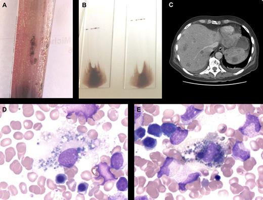
A 60-year-old man was hospitalized for weakness, back pain, and respiratory distress. Multiple lymph nodes, liver, and spleen enlargements were palpable. Blood cell count findings were hemoglobin 9.4 g/dL, white blood cells 7.910 × 10⁹/L, and platelets 6 × 10⁹/L. Peripheral blood film observation revealed leuco-erythroblastosis. A bone marrow aspiration yielded a viscous mixture with brownish clumps (see figure panel A) leading to dark smears (panel B). The marrow was heavily infiltrated by noncohesive tumor cells, and the cytoplasm contained heterogeneous dark brown granules (panel D). Numerous background macrophages also showed cytoplasmic pigmentation (melanophages, panel E). Bone marrow biopsy immunochemistry was positive for Melan A. A dorsal melanoma was found, as well as widespread vertebral, liver, and spleen metastases (panel C). Despite intensive care the patient died.
Metastatic melanoma involving the bone marrow is rare, occurring in 5% of patients with disseminated disease but in up to 45% when an autopsy-staging procedure is performed. Dark pigments are highly suggestive of melanoma cells and are often found in the associated macrophages. Although the diagnosis should be relatively straightforward, the nature of the pigment should be confirmed using melanoma-associated MART-1 gene product (Melan A, A 103 antibody) or melanosome matrix protein gp 100 (HMB 45 antibody).
For additional images, visit the ASH IMAGE BANK, a reference and teaching tool that is continually updated with new atlas and case study images. For more information visit http://imagebank.hematology.org.


This feature is available to Subscribers Only
Sign In or Create an Account Close Modal