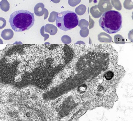
A 77-year-old man with seropositive rheumatoid arthritis was investigated for mild pancytopenia. He had mild splenomegaly, but no adenopathy. Hemoglobin was 117 g/L, platelets 126 × 109/L, and leukocytes 2.0 × 10⁹/L with 96% lymphocytes, most of which were large granular lymphocytes (LGLs). Immunophenotyping showed an aberrant T-cell population co-expressing CD3, CD8, and CD57 with reduced expression of CD5, and absent CD56. T-cell receptor gene rearrangement confirmed a monoclonal T-cell population, consistent with T-LGL leukemia. After treatment with methotrexate lymphocytosis and splenomegaly resolved, but cytoplasmic inclusions remained. Approximately 40% of lymphocytes contained cytoplasmic inclusions, either single or multiple, and in some cells the inclusions were associated with a cytoplasmic vacuole (see figure). The inclusions had a color resembling that of nuclear chromatin, with variable shapes ranging from round to rod-like. Electron microscopy showed them as giant parallel tubular arrays (PTAs) consisting of dense bundles of thick-walled tubules with a diameter of approximately 30 nm.
First recognized more than 40 years ago, the function of these structures is unclear. PTAs can be found in infectious mononucleosis, and rarely in chronic lymphocytic leukemia and in healthy individuals. Most often they are seen in systemic immunologic disorders (eg, systemic lupus erythematosus, rheumatoid arthritis, HIV infection), all of which can be associated with reactive or clonal proliferation of T-LGLs.
For additional images, visit the ASH IMAGE BANK, a reference and teaching tool that iscontinually updated with new atlas and case study images. For more information visit http://imagebank.hematology.org.


This feature is available to Subscribers Only
Sign In or Create an Account Close Modal