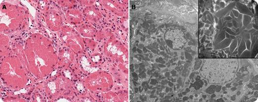
A 67-year-old man with an elevated creatinine (231 mmol/L) was found to have an elevated IgG monoclonal gammopathy. A renal biopsy identified light chain deposition. Routine staining by hematoxylin and eosin stain revealed acute tubular necrosis, with tubular dilatation, sloughing of the epithelial cells, loss of nuclei, and cytoplasmic fragmentation (panel A; original magnification ×400). On electron microscopy, there were the typical abundant needle-like crystalline materials in the tubular epithelial cells (panel B; original magnification ×1650, original inset magnification ×7300). Complete blood count at baseline revealed the following: white blood cells, 10.6 × 10⁹/L; hemoglobin, 135 g/L; mean corpuscular volume, 90.6 fL; platelets, 233 × 109/L. Calcium, albumin, and a skeletal survey were normal. A bone marrow aspirate and biopsy confirmed an underlying diagnosis of multiple myeloma as the cause of renal light chain deposition. He was started on cyclophosphamide, bortezomib, and dexamethasone, followed by autologous stem cell transplant.
Light chain deposition disease is a rare type of plasma cell dyscrasia characterized by deposition of immunoglobulin fragments mostly involving the kidneys, but it may affect other organs such as the heart or liver. Concurrent multiple myeloma may be present in half of the cases. Renal biopsy often shows linear deposits of monoclonal light chains along the tubular basement membrane. Unlike amyloidosis, light chains in light chain deposition disease do not undergo conformational changes to form fibrils and do not have the ability to bind Congo Red.
For additional images, visit the ASH IMAGE BANK, a reference and teaching tool that is continually updated with new atlas and case study images. For more information visit http://imagebank.hematology.org.


This feature is available to Subscribers Only
Sign In or Create an Account Close Modal