Key Points
Exosomal NKp30-ligand BAG6 is crucial for detection of tumor cells by NK cells in vitro and in vivo.
Soluble plasma factors including BAG6 suppress NK cell cytotoxicity and promote evasion of CLL cells from NK cell anti-tumor activity.
Abstract
Natural killer (NK) cells are a major component of the anti-tumor immune response. NK cell dysfunctions have been reported in various hematologic malignancies, including chronic lymphocytic leukemia (CLL). Here we investigated the role of tumor cell-released soluble and exosomal ligands for NK cell receptors that modulate NK cell activity. Soluble CLL plasma factors suppressed NK cell cytotoxicity and down-regulated the surface receptors CD16 and CD56 on NK cells of healthy donors. The inhibition of NK cell cytotoxicity was attributed to the soluble ligand BAG6/BAT3 that engages the activating receptor NKp30 expressed on NK cells. Soluble BAG6 was detectable in the plasma of CLL patients, with the highest levels at the advanced disease stages. In contrast, NK cells were activated when BAG6 was presented on the surface of exosomes. The latter form was induced in non-CLL cells by cellular stress via an nSmase2-dependent pathway. Such cells were eliminated by lymphocytes in a xenograft tumor model in vivo. Here, exosomal BAG6 was essential for tumor cell killing because BAG6-deficient cells evaded immune detection. Taken together, the findings show that the dysregulated balance of exosomal vs soluble BAG6 expression may cause immune evasion of CLL cells.
Introduction
Chronic lymphocytic leukemia (CLL) patients suffer from severe immune defects resulting in increased susceptibility to infections and failure to generate an anti-tumor immune response.1 Natural killer (NK) cells, lymphocytes of the innate immune system, are considered to be a major component of the immunosurveillance in leukemia.2-4 However, little is known about the functionality of NK cells and their role in tumor immune escape in CLL.
NK cells are tightly regulated by inhibitory or activating “missing self” and “induced self” signals sensed via cell surface receptors.5 The best examined activating receptors are the Fc receptor CD16, NKG2D, and the natural cytotoxicity receptors (NCRs) NKp30, NKp44, and NKp46. Known ligands for NKG2D are the major histocompatibility complex (MHC) class I-related molecules MICA/B and the UL16-binding proteins (ULBP1, ULBP2, ULBP3, ULBP4, ULBP5, and ULBP6) that are induced upon cellular stress on target cells.6,7 Only a few ligands for the NCRs have been identified to date.8-14 Surprisingly, among novel ligands for NKp30 (BAG6 [BAT3],10 B7-H611 ), NKp44 (proliferating cell nuclear antigen12 ) and NKp46 (vimentin13,14 ), only B7-H6 is a surface membrane ligand. BAG6, proliferating cell nuclear antigen, and vimentin are proteins without any classical transmembrane domain and are known to exert divergent intracellular functions, including protein sorting and transport, proliferation, and apoptosis. It is still not clear how these intracellular proteins are exposed to surface NK cell receptors.
Recently it was shown that NK cells display a poor cytolytic activity against CLL cells, which could be restored with IL-2/IL-15,15 but the mechanisms for NK cell suppression or anergy remain to be elucidated. The NKG2D expression on NK cells in CLL was not significantly altered in comparison with healthy donors,15-17 although it was reported that CLL patients have high serum levels of soluble NKG2D ligands. Shedding of NKG2D ligands from the surface of tumor cells represents an evasion strategy to escape from NK cell-mediated recognition and killing in hematologic and solid tumors.18,19 Among the activating NK cell receptors, only NKp30 expression was significantly reduced on NK cells in CLL patients.15 This is interesting because NKp30 is a receptor not only involved in direct target cell killing but it is also responsible for the interaction with dendritic cells that represents the link to the adaptive immune response.21-23 Under certain conditions and by mechanisms that are not completely understood, BAG6 can be released from cells into the extracellular environment.10,24,25 The protein can be expressed on the surface of exosomes to engage NKp30 and to activate NK cells.10,25 Exosomes are 50 to 100 nm microvesicles that originate from intracellular multivesicular bodies and are produced by many cell types.26,27 The inducible formation and release of exosomes depends on the DNA damage-induced p53-dependent secretory pathway.28-30
To address the role of NKp30 and its ligand BAG6 for immunosurveillance in CLL, we analyzed the release of BAG6 from tumor cell lines and CLL cells, and the BAG6-dependent modulation of NK cell activity. We demonstrate in vitro and in vivo that BAG6 plays an important role in recognition and killing of tumor cells by NK cells and provides a possible explanation for the reduced efficacy of NK cells in CLL patients.
Materials and methods
Human samples
The collection of and the experiments with human plasma, blood samples of CLL patients, and healthy donors were approved by the local ethics committee of the University of Cologne under reference numbers 08-275 and 11-140, and donors provided written consent in accordance with the Declaration of Helsinki. Plasma was diluted 1:1 with phosphate-buffered saline (PBS), and stored at −80°C.
Cells
NK cells were purified from peripheral blood mononuclear cells (PBMCs) with the human NK cell isolation kit using an autoMACS Pro Separator (Miltenyi, Bergisch-Gladbach, Germany), according to the manufacturer’s instruction. Primary lymphocytes were cultured in Iscove modified Dulbecco medium supplemented with 10% fetal bovine serum and antibiotics. The human embryonic kidney line 293T (ACC-635), the breast cancer line MCF7 (ACC-115), the Burkitt lymphoma line Raji (ACC-319), the B-cell precursor leukemia lines NALM6 (ACC-128), and 697 (ACC-42) were purchased from the DSMZ (Braunschweig, Germany) and cultured, as recommended. We also used the chronic B-cell leukemia cell lines JVM-3 (ACC-18, p53 wild type [wt]) and MEC-1 (ACC-497, p53 mutated).
ELISA
The mouse monoclonal 3E4 (raised against the BAG6 N-terminus) and the BAG6-specific rabbit serum 416010 were used to detect BAG6. MICA, MICB, migration inhibitory factor, and ULBP2 were measured with commercial enzyme-linked immunosorbent assay (ELISA) kits (R&D Systems, Wiesbaden, Germany). Details of protocol deviations are given in the supplements.
Flow cytometry
The following reagents were used: mouse anti-human CD9, monoclonal mouse anti-human-BAG6, and annexinV-phycoerythrin (PE). As a secondary antibody, PE goat anti-mouse was used. Binding of recombinant NKp30-Fc protein was detected by Cy3 coupled anti-human Fc.
Primary NK cells were stained with fluorescein isothiocyanate- or PE-labeled antigen-specific antibodies against NKG2D, NKp30, NKp44, NKp46, CD16, CD56, and the activation marker CD69, and were analyzed using a fluorescence-activated cell sorter (FASC) Calibur (Beckton Dickinson, Heidelberg, Germany) with FlowJo software (Treestar, Ashland, OR). For antibody details, please see the supplement to this article.
Transgenic cells
MCF7 cells overexpressing nSmase2 were previously described.31 Knockdown of nSmase2 was achieved using specific small-interfering RNA (siRNA) (QIAGEN, Hilden, Germany). The lentiviral particle production and transduction of 293T cells for BAG6 knockdown were achieved with the Mission shRNA Vector pLKO.1-puro System using shRNA Plasmid TRC7354 (Sigma-Aldrich, Munich, Germany). Experimental procedures were carried out according to the manufacturers’ instructions. BAG6-GFP-fusion sequences were cloned into the vector pEGFP-C1 (Clontech, Mountain View, CA).
Cytotoxicity assays
NK cells were left untreated or pre-incubated for 20 to 48 hours with 25% serum in Iscove modified Dulbecco medium, recombinant BAG6 (10 ng/mL, Abcam) or with exosomes purified from 293T cells prior standard cytotoxicity assays.10 Blocking antibodies or Fc constructs were added as indicated (10 µg/mL).
Exosome preparation
Exosome production, purification via centrifugation and the coupling to beads for flow cytometry was previously described.25 In brief, cells were cultured in media with exosome-depleted fetal calf serum (100 000 g for 90 minutes). The supernatant was centrifuged for 10 minutes at 300 g and twice for 15 minutes at 6800 g to remove cell fragments. Exosomes were pelleted twice at 100 000 g for 90 minutes with intermediate resuspension in PBS. The amount of exosomal protein was quantified by Nanodrop1000 (Thermo Scientific, Schwerte, Germany). Plasma samples were diluted 1:1 in PBS prior to exosome purification.
Western blot
Cells were lyzed in lysis buffer (50 mM Tris-HCl pH 8, 150 mM NaCl, 0.5% Triton X-100, 0.5% protease inhibitors). Protein concentration was measured with a protein assay. Then 10 to 30 µg were loaded onto sodium dodecyl sulfate-polyacrylamide gel electrophoresis gels for blotting using standard methods (see supplemental Table 1).
In vivo experiments
The (3 × 10⁶) 293T cells were transplanted subcutaneously into B-, T-, and NK cell-deficient severe combined immunodeficiency (SCID)/beige mice (Charles River Laboratories, Sulzfeld, Germany) followed by PBMC injections (3 × 10⁶ cells intraperitoneally). Tumor growth was monitored with calipers and tumor volume calculated as length × breadth × height/2. Animal experiments were approved from local authorities (State Northrhine-Westfalia, Landesamt für Natur, Umwelt und Verbraucherschutz, ethical approval no. 31.07.135).
Statistical analysis
Data were analyzed with GraphPad Prism 5 (San Diego, CA) and appropriate statistical tests were chosen as indicated in the figure legends. Bar diagrams are presented as mean plus SEM.
Results
Tumor cell lines release BAG6 via nSmase2-dependent exosomal pathway
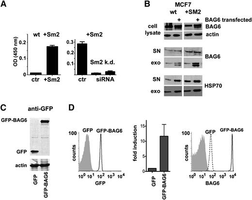
We investigated the exosomal pathway responsible for the BAG6 release in MCF7 cells. We chose MCF7 cells as the model, as these are p53-proficient and characterized by low endogenous expression of the tumor necrosis factor (TNF)-α-inducible enzyme neutral sphingomyelinase 2 (nSmase2/SMPD3). Both p53 and nSmase2 are mandatory for exosome formation.28-30,32 Here overexpression of nSmase2 resulted in a robust release of BAG6-expressing exosomes, which were not detectable in the parental wt cell line (Figure 1A, left panel). The elevated release could be suppressed upon down-regulation of the transgene with 2 different siRNAs targeting nSmase2 (Figure 1A, right panel). Western blot analysis (Figure 1B) of exosomes from wt MCF7 cells, which were transfected with BAG6, revealed increased BAG6 expression in the exosomal fraction. The expression of HSP70, known as an exosomal marker and a BAG6-interacting protein,25 was enhanced in both the crude cell supernatant and in the exosomal lysate. This effect was even more pronounced in nSmase2-overexpressing MCF7 cells upon transfection with BAG6 (Figure 1B), together indicating that BAG6 was released via the exosomal pathway in an nSmase2-dependent manner.
Tumor cell lines release BAG6 via the exosomal pathway. (A) Exosomes were isolated from MCF7 wt cells, from MCF7 cells stably overexpressing Smase2 (+Sm2) (left panel) and from MCF7+Sm2 cells upon control siRNA (AF488) transfection or siRNA-mediated knockdown of Smase2 (right panel) to detect BAG6 on exosomes by ELISA. Bar diagrams show mean and SEM (n = 3). See supplemental Figure 1 for reverse transcription-polymerase chain reaction to detect nSmase2 mRNA in wt cells or upon overexpression/down-regulation of nSmase2, respectively. (B) Western blots to detect BAG6, actin, and HSP70 in cell lysates, supernatant (SN) and purified exosomes (exo) from wt and +Sm2-MCF7 cells upon control and BAG6-transfection. (C) Western blot of 293T lysates with an anti-GFP antibody to detect expression of transfected GFP (control plasmid) or GFP-BAG6 fusion protein, consisting of GFP and BAG6 full length protein. Actin was used as a loading control. (D) FASC analysis of exosomes. The exosomes were purified from transfected cells and GFP-expressing exosomes were detected by GFP-fluorescence (left histogram): gray-filled histogram, GFP; black line, GFP-BAG6. Right histogram: anti-BAG6 staining of the same exosomes, detecting transfected and endogenous BAG6. Gray-filled histogram, istoype antibody; dashed line, GFP; thick black line, GFP-BAG6. GFP-expression data of 7 transfections were summarized in a bar diagram (mean and standard error of the mean).The means are significantly different (P = .0005, Wilcoxon signed-rank test).
Tumor cell lines release BAG6 via the exosomal pathway. (A) Exosomes were isolated from MCF7 wt cells, from MCF7 cells stably overexpressing Smase2 (+Sm2) (left panel) and from MCF7+Sm2 cells upon control siRNA (AF488) transfection or siRNA-mediated knockdown of Smase2 (right panel) to detect BAG6 on exosomes by ELISA. Bar diagrams show mean and SEM (n = 3). See supplemental Figure 1 for reverse transcription-polymerase chain reaction to detect nSmase2 mRNA in wt cells or upon overexpression/down-regulation of nSmase2, respectively. (B) Western blots to detect BAG6, actin, and HSP70 in cell lysates, supernatant (SN) and purified exosomes (exo) from wt and +Sm2-MCF7 cells upon control and BAG6-transfection. (C) Western blot of 293T lysates with an anti-GFP antibody to detect expression of transfected GFP (control plasmid) or GFP-BAG6 fusion protein, consisting of GFP and BAG6 full length protein. Actin was used as a loading control. (D) FASC analysis of exosomes. The exosomes were purified from transfected cells and GFP-expressing exosomes were detected by GFP-fluorescence (left histogram): gray-filled histogram, GFP; black line, GFP-BAG6. Right histogram: anti-BAG6 staining of the same exosomes, detecting transfected and endogenous BAG6. Gray-filled histogram, istoype antibody; dashed line, GFP; thick black line, GFP-BAG6. GFP-expression data of 7 transfections were summarized in a bar diagram (mean and standard error of the mean).The means are significantly different (P = .0005, Wilcoxon signed-rank test).
Next, BAG6 full length protein was fused to a GFP reporter gene to test whether the expression of BAG6 on exosomes was the result of an active translocation. The GFP control and the GFP-BAG6 fusion protein were expressed at comparable levels in transfected 293T cells (Figure 1C). Exosomes were purified and coupled with beads to analyze GFP-fluorescence and BAG6 expression by flow cytometry (Figure 1D). Exosomes from GFP-transfected cells showed background fluorescence (same level as untransfected cells), whereas a clear targeting of GFP to exosomes was observed for exosomes from GFP-BAG6 transfected cells indicating that BAG6 contains exosome-targeting sequences (see bar diagram for quantitative results). In line, we observed an increase of BAG6 expression on exosomes upon GFP-BAG6 transfection (Figure 1D, right histogram). To exclude potential interference of the GFP tag with the subcellular distribution and exosomal targeting of the proteins we performed analogous experiments with 6 × his-tagged constructs that confirmed the above data (not shown).
BAG6-expressing exosomes trigger NK cell cytotoxicity
Having established that BAG6 is released via exosomes in vitro, we were interested in the functional activity of BAG6-expressing exosomes. Because it is known that exosomes are released in response to the DNA damage-inducible p53-dependent secretory pathway,28-30 we first tested whether the release of BAG6-expressing exosomes was inducible from cells with an intact p53 pathway (293T cells). Exosomal BAG6 release was triggered in response to cellular stress signals (heat shock or doxorubicine) or upon inflammatory signals including TNF-α or HDAC inhibition with valproate (Figure 2A). Therefore, 293T cells were used to produce exosomes for NK cell stimulation. The overexpression of BAG6 on exosomes (as previously shown25 ) was confirmed here by flow cytometry of the exosomes using a BAG6-specific antibody (Figure 2B) and a recombinant NKp30-Fc (data not shown). Exosomal marker proteins were detectable in exosomal lysates from wt and BAG6-overexpressing cells, however, due to the detection threshold, BAG6 was only detectable by Western blot analysis in exosomes from BAG6-transfected cells (Figure 2C). Healthy NK cells were incubated with exosomes derived from wt or BAG6 overexpressing cells and used in killing assays with the leukemic cell lines 697 and NALM6 as target cells (Figure 2D). Exosome-treated NK cells were able to kill these target cells that were initially resistant against NK cell lysis. The killing efficacy of NK cells treated with BAG6-overexpressing exosomes was even higher compared with NK cells treated with wt exosomes and could be blocked with an NKp30 antibody (Figure 2D).
BAG6-positive exosomes trigger NK cell cytotoxicity. (A) BAG6 was detected on exosomes released by 293T cells either constitutively or in response to cellular stress, including treatment with heat shock (HS, 30 minutes, 42°C and 2 hours recovery), doxorubicine (50 nM), TNF-α (50 ng/mL), and valproate (100 μg/mL). Mean and standard error of the mean (SEM) are indicated (n = 3), the enhanced BAG6 release was significant compared with untreated cells (HS: P = .0158; doxo: P = .0142, TNF-α: 0.0037, valproate: P = .001, unpaired Student t test). (B) Flow cytometry demonstrated binding of a BAG6-specific antibody (3E4) and a CD9-specific antibody to exosomes from wt cells (dotted line, endosomes) and exosomes from BAG6-transfected cells (+BAG6, black line). Gray-filled histogram: isotype control. FACS, fluorescence-activated cell sorter. (C) Western blot (WB) analysis to detect BAG6 and exosome marker (HSP70, CD9, CD81, CD63) in exosomes lysates of wt and BAG6-transfected cells (+BAG6). (D) BAG6-expressing exosomes enhanced the NK cell killing activity in cytotoxicity assays against the NK cell-resistant target cell lines 697 and NALM6 because untreated NK cells were less cytolytic than NK cells treated with exosomes purified from wt 293T cells (+exo). NK cells treated with exosomes from BAG6-transfected 293T cells (+BAG6-exo) demonstrated the highest killing activity. One representative of 3 experiments is shown. Blocking of NKp30 (clone P30-15 and IgG1-isotype control from BioLegend) reduced the target cell killing significantly (mean and SEM [n = 3]); 697: P = .0200 and Nalm6: P = .0123, unpaired Student t test; effector:target ratio, 10:1).
BAG6-positive exosomes trigger NK cell cytotoxicity. (A) BAG6 was detected on exosomes released by 293T cells either constitutively or in response to cellular stress, including treatment with heat shock (HS, 30 minutes, 42°C and 2 hours recovery), doxorubicine (50 nM), TNF-α (50 ng/mL), and valproate (100 μg/mL). Mean and standard error of the mean (SEM) are indicated (n = 3), the enhanced BAG6 release was significant compared with untreated cells (HS: P = .0158; doxo: P = .0142, TNF-α: 0.0037, valproate: P = .001, unpaired Student t test). (B) Flow cytometry demonstrated binding of a BAG6-specific antibody (3E4) and a CD9-specific antibody to exosomes from wt cells (dotted line, endosomes) and exosomes from BAG6-transfected cells (+BAG6, black line). Gray-filled histogram: isotype control. FACS, fluorescence-activated cell sorter. (C) Western blot (WB) analysis to detect BAG6 and exosome marker (HSP70, CD9, CD81, CD63) in exosomes lysates of wt and BAG6-transfected cells (+BAG6). (D) BAG6-expressing exosomes enhanced the NK cell killing activity in cytotoxicity assays against the NK cell-resistant target cell lines 697 and NALM6 because untreated NK cells were less cytolytic than NK cells treated with exosomes purified from wt 293T cells (+exo). NK cells treated with exosomes from BAG6-transfected 293T cells (+BAG6-exo) demonstrated the highest killing activity. One representative of 3 experiments is shown. Blocking of NKp30 (clone P30-15 and IgG1-isotype control from BioLegend) reduced the target cell killing significantly (mean and SEM [n = 3]); 697: P = .0200 and Nalm6: P = .0123, unpaired Student t test; effector:target ratio, 10:1).
BAG6 is mandatory for immune surveillance in a 293T xenograft model
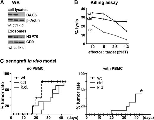
Next we investigated the loss of function of BAG6 on exosomes in NK cell-mediated killing in vitro and for immune surveillance in vivo. To this end, we established a BAG6-deficient 293T cell line using a short hairpin RNA (shRNA)-mediated knockdown (Figure 3A). The exosomal markers CD9 and HSP70 were still expressed in the exosomal fractions of BAG6 knockdown (k.d.) cells compared with fractions of wt or control transduced cells (ctrl), when equal protein concentrations were loaded (Figure 3A). The protein amount of the exosomal fraction normalized to the cell number used was also similar between the cell lines (not shown). Knockdown of BAG6 did not result in substantial changes of surface molecules on cells and released exosomes such as MHC I, MHC II, intercellular adhesion molecule-1, NKG2D ligands, and NKp46 ligands, except BAG6, which was not expressed on k.d. cells and exosomes (supplemental Figure 2). Next, an NK cell killing assay using 293T wt and transduced cells was performed. As expected, and as previously described for other cell types,10,12,25 target cells without BAG6 expression were more resistant against NK cell killing than parental wt cells or shRNA (Figure 3B).
Role of BAG6 for immune detection in vitro and in an in vivo xenograft model. (A) Western blot (WB) of 293T cell lysates to verify BAG6 downregulation in BAG6-shRNA–transduced 293T cells (k.d.) in comparison with wt and control shRNA-transduced cells (ctrl). Actin was used as a loading control. Exosomes derived from these cell lines were checked for exosomal markers HSP70 and CD9. BAG6 knockdown in exosomes was demonstrated via flow cytometry (supplementary Figure 2). (B) The wt, control shRNA (ctrl), and BAG6 shRNA (k.d.) transduced 293T cells were used as target cells in an NK cell cytotoxicity assay (1 representative experiment of 3). (C) The wt, ctrl, and k.d. 293T cells were implanted subcutaneously (3 × 10⁶ cells) into immunodeficient SCID/beige mice. The treatment groups received human lymphocytes (1 day after tumor cell transplantation, 3 × 10⁶ PBMC intraperitoneally.). The tumor incidence was monitored (left: no PBMCs; right: with PBMCs; n = 9 or 10 for ctrl and k.d.; n = 5 for wt in both settings).
Role of BAG6 for immune detection in vitro and in an in vivo xenograft model. (A) Western blot (WB) of 293T cell lysates to verify BAG6 downregulation in BAG6-shRNA–transduced 293T cells (k.d.) in comparison with wt and control shRNA-transduced cells (ctrl). Actin was used as a loading control. Exosomes derived from these cell lines were checked for exosomal markers HSP70 and CD9. BAG6 knockdown in exosomes was demonstrated via flow cytometry (supplementary Figure 2). (B) The wt, control shRNA (ctrl), and BAG6 shRNA (k.d.) transduced 293T cells were used as target cells in an NK cell cytotoxicity assay (1 representative experiment of 3). (C) The wt, ctrl, and k.d. 293T cells were implanted subcutaneously (3 × 10⁶ cells) into immunodeficient SCID/beige mice. The treatment groups received human lymphocytes (1 day after tumor cell transplantation, 3 × 10⁶ PBMC intraperitoneally.). The tumor incidence was monitored (left: no PBMCs; right: with PBMCs; n = 9 or 10 for ctrl and k.d.; n = 5 for wt in both settings).
All 3 of the 293T cell lines (wt, ctrl, k.d.) were able to form tumors upon subcutaneous injection in SCID/beige mice deficient for B-, T-, and NK cells, although tumor formation of the transduced cell lines was slightly delayed (Figure 3C). This might reflect the slightly reduced proliferation rate of the transduced cells that we observed in vitro (not shown). The mice received either human PBMCs or PBS control injections 1 day after tumor cell inoculation. Only the wt and the ctrl lines were efficiently recognized and eliminated by PBMCs, whereas the BAG6 knockdown cell line was able to form tumors, despite the presence of immune cells, albeit at a slightly lesser incidence than PBS-injected animals but nevertheless statistically significant (Figure 3C and supplemental Figure 3). Hence, the BAG6 knockdown triggered escape from immunologic tumor surveillance demonstrating the role of exosomal BAG6 immunosurveillance in vivo.
Elevated plasma levels of NKp30-ligand BAG6 and ligands for NKG2D in CLL patients
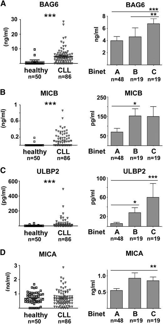
A defective cytolytic activity of NK cells has been recently described in patients with CLL.15,16 To assess the role of BAG6 for NK cell activity in this disease, we measured BAG6 plasma levels in 86 primary CLL samples and in 50 age-matched healthy controls with ELISA. Surprisingly, CLL patients had significantly elevated levels of BAG6 compared with healthy donors. Moreover, BAG6 level increased with progressed disease stage (Figure 4A). Because a role for soluble NKG2D ligands in CLL is discussed,16,17 in our analysis we included ligands for the activating NK cell receptor NKG2D (MICB, ULBP2, and MICA). Interestingly, we found significantly higher levels of MICB and ULBP2 in patients and levels increased with advanced disease stage (Figure 4B-C). MICA was already discussed as a prognostic factor in CLL.17 However, we did not measure any significant overall difference in the expression levels of MICA in healthy vs CLL samples, although MICA plasma levels were significantly higher in CLL patients with advanced Binet stage C compared with Binet stage A (Figure 4D). Taken together, patients with CLL showed elevated levels of soluble ligands for NKp30 and NKG2D in comparison with healthy individuals.
Plasma levels of BAG6 and NKG2D ligands are elevated in CLL patients and increase with advanced Binet stage. (A-D) Plasma levels of NKp30-ligand BAG6 and NKG2D ligands (MICB, ULBP2, MICA) were measured by ELISA in healthy controls (age-matched) and CLL samples. A comparison of healthy vs CLL samples and a comparison of CLL samples (mean and standard error of the mean) from patients with Binet stage A, B, or C are depicted. Significant differences are indicated (two-tailed, unpaired Mann-Whitney U test).
Plasma levels of BAG6 and NKG2D ligands are elevated in CLL patients and increase with advanced Binet stage. (A-D) Plasma levels of NKp30-ligand BAG6 and NKG2D ligands (MICB, ULBP2, MICA) were measured by ELISA in healthy controls (age-matched) and CLL samples. A comparison of healthy vs CLL samples and a comparison of CLL samples (mean and standard error of the mean) from patients with Binet stage A, B, or C are depicted. Significant differences are indicated (two-tailed, unpaired Mann-Whitney U test).
Soluble plasma factors from CLL patients impair NK cell function
In cytotoxicity assays, the natural cytotoxic activity of NK cells is believed to mainly depend on NKG2D and NCRs that synergize with CD16.33 Having identified BAG6 as an extracellular ligand of the NCR NKp30 that activates NK cells10 (Figure 2) and promotes immunosurveillance (Figure 3), the overexpression of BAG6 in the plasma of CLL patients with impaired NK cell activity was quite unexpected. Hence, we tested the impact of plasma from CLL patients on natural cytotoxicity of NK cells. To this end, NK cells were purified from healthy donors and incubated overnight with plasma derived from CLL patients or age-matched healthy volunteers. The cytotoxicity of plasma-treated and untreated NK cells (control) was assessed in killing assays using the Burkitt’s lymphoma cell line Raji as target. Samples from healthy donors had only minor effects on NK cell cytotoxicity, whereas killing activity was eliminated upon incubation with CLL plasma (Figure 5A). Flow cytometry (Figure 5B) revealed that the activating receptors NKp30, NKp46, and NKG2D were not significantly modulated, although both, healthy-donor and CLL samples, induced a minor down-regulation of these receptors. Instead, a robust down-regulation of surface CD16 and CD56 was observed upon treatment of normal NK cells with CLL sera (Figure 5B). Control experiments confirmed that the reduced CD16 expression was not due to a blocking of antibody binding by serum IgG (data not shown). Next, the plasma samples were fractionated into the soluble and vesicular part to test which of these fractions induced the NK cell inhibition. As expected, the different fractions of healthy samples did not alter the cytotoxicity of NK cells significantly, whereas the CLL plasma-dependent inhibition was mainly mediated by soluble factors but not by the exosomal fraction (Figure 5C). This is in line with our previous studies that reported recombinant, purified BAG6 inhibited TNF-α and IFN-γ release from NK cells,10 as well as NK cell cytotoxicity.25 Collecting data from patients (n = 6) (Figure 5D) revealed that the inhibition mediated by plasma and the soluble fractions was significant, whereas the exosomal fractions rather enhanced cytotoxicity, albeit the differences to the untreated control, were minor and not significant.
Plasma and the soluble plasma fraction of CLL patients inhibit NK cell function. (A) Cytotoxicity assays using NK cells of healthy donors that were left untreated (control) or incubated overnight with 25% plasma of healthy donors (n = 3) or CLL patients (n = 3). The ratio of effector NK cells to target cells (Raji, B lymphocyte cell line) is indicated. (B) NK cell markers and activating receptors expressed on control NK cells (incubated in medium) and NK cells that were incubated with healthy donor or CLL patient plasma were analyzed by flow cytometry. A significant down-regulation was observed for CD16 and CD56 upon incubation with CLL plasma (n = 3 for donor NK cells; n = 3 or 6 for plasma from healthy donors or CLL patients). (C) NK cells were left untreated (control), treated with plasma samples (pl), the soluble plasma fraction (so), or the exosomal/vesicular fraction (exo) derived from healthy donors or CLL patients and then used for a cytotoxicity assay. One representative example is shown (left); the bar diagram (D) summarizes data from 6 CLL samples. The inhibition of NK cell cytotoxicity by CLL plasma or the soluble plasma fraction was significant (n = 6).
Plasma and the soluble plasma fraction of CLL patients inhibit NK cell function. (A) Cytotoxicity assays using NK cells of healthy donors that were left untreated (control) or incubated overnight with 25% plasma of healthy donors (n = 3) or CLL patients (n = 3). The ratio of effector NK cells to target cells (Raji, B lymphocyte cell line) is indicated. (B) NK cell markers and activating receptors expressed on control NK cells (incubated in medium) and NK cells that were incubated with healthy donor or CLL patient plasma were analyzed by flow cytometry. A significant down-regulation was observed for CD16 and CD56 upon incubation with CLL plasma (n = 3 for donor NK cells; n = 3 or 6 for plasma from healthy donors or CLL patients). (C) NK cells were left untreated (control), treated with plasma samples (pl), the soluble plasma fraction (so), or the exosomal/vesicular fraction (exo) derived from healthy donors or CLL patients and then used for a cytotoxicity assay. One representative example is shown (left); the bar diagram (D) summarizes data from 6 CLL samples. The inhibition of NK cell cytotoxicity by CLL plasma or the soluble plasma fraction was significant (n = 6).
BAG6 is released from CLL cells and cell lines as a soluble factor and suppresses NK cell cytotoxicity
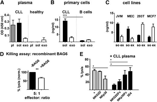
To understand a possible contribution of BAG6 to NK cell inhibition in CLL, we analyzed the distribution of BAG6 in soluble vs exosomal fractions in plasma of CLL patients (Figure 6A). In healthy donor samples, BAG6 was barely detectable and the protein was sequestered within the exosomal fraction. In marked contrast, most of the BAG6 protein accumulated in the soluble fraction of CLL patient plasma (Figure 6A). BAG6 was also detectable in the soluble part of supernatant from suspension-cultured CLL cells, but barely in association with the exosomal fraction. The same amount of cultured B cells purified from healthy donors did not release much BAG6 (Figure 6B). The fractionation of the supernatant from CLL cell lines JVM-3 and MEC-1 revealed a similar distribution with high levels of the soluble protein. However, 293T cells and MCF7 cells released the exosomal form (Figure 2B and Figure 6C).
BAG6 is released from CLL cells and cell lines as a soluble factor and inhibits NK cell cytotoxicity. (A) BAG6 was quantified in plasma (pl), soluble (so), and exosomal (exo) fraction of healthy donors and CLL patients using ELISA. Mean and standard error of the mean (SEM) from patients (n = 6) or healthy donors (n = 3) are depicted. (B) BAG6 ELISA of the soluble (so) and exosomal (exo) fraction of supernatant derived from cultured primary CLL or healthy B cells. Mean and SEM of 4 experiments are indicated. The amount of soluble BAG6 is significantly higher in CLL-derived supernatant in comparison with healthy B cell supernatant (P = .0001). The differences among the exosomal fractions are not significant (P = .0967); unpaired Student t test. (C) BAG6 concentration in fractionated supernatants of the CLL cell lines JVM-3, MEC-1, the human embryonic kidney cell line 293T, and the breast cancer cell line MCF7 was determined with ELISA. The differences of BAG6 distribution in the soluble vs the exosomal fraction are significant (mean and SEM, n = 4) between JVM/MEC in comparison with 293T/MCF7; unpaired Student t test. (D) NK cells were incubated with healthy donor plasma with or without soluble BAG6 protein (10 ng/mL) and their cytotoxicity was assessed in an NK cell cytotoxicity assay using the target cell line Raji. Normalized lysis is shown (mean and SEM of 4 independent experiments). (E) NK cells were incubated with control (gray bars) or CLL patient plasma (25% overnight, black bars) and the cytotoxicity against Raji cells (effector:target ratio, 10:1) was measured in the presence of isotype antibody (-), NKG2D blocking antibody (clone 1D11), NKp30 blocking antibody (clone P30-15), Fc control protein (-), NKG2D-Fc, or NKp30-Fc. NK cells were incubated with blocking constructs (10 μg/mL). Depletion of BAG6 from CLL patient plasma was performed using the monoclonal 3E4 antibody. Mean and SEM of 3 independent experiments are shown; significant differences are indicated; unpaired Student t test.
BAG6 is released from CLL cells and cell lines as a soluble factor and inhibits NK cell cytotoxicity. (A) BAG6 was quantified in plasma (pl), soluble (so), and exosomal (exo) fraction of healthy donors and CLL patients using ELISA. Mean and standard error of the mean (SEM) from patients (n = 6) or healthy donors (n = 3) are depicted. (B) BAG6 ELISA of the soluble (so) and exosomal (exo) fraction of supernatant derived from cultured primary CLL or healthy B cells. Mean and SEM of 4 experiments are indicated. The amount of soluble BAG6 is significantly higher in CLL-derived supernatant in comparison with healthy B cell supernatant (P = .0001). The differences among the exosomal fractions are not significant (P = .0967); unpaired Student t test. (C) BAG6 concentration in fractionated supernatants of the CLL cell lines JVM-3, MEC-1, the human embryonic kidney cell line 293T, and the breast cancer cell line MCF7 was determined with ELISA. The differences of BAG6 distribution in the soluble vs the exosomal fraction are significant (mean and SEM, n = 4) between JVM/MEC in comparison with 293T/MCF7; unpaired Student t test. (D) NK cells were incubated with healthy donor plasma with or without soluble BAG6 protein (10 ng/mL) and their cytotoxicity was assessed in an NK cell cytotoxicity assay using the target cell line Raji. Normalized lysis is shown (mean and SEM of 4 independent experiments). (E) NK cells were incubated with control (gray bars) or CLL patient plasma (25% overnight, black bars) and the cytotoxicity against Raji cells (effector:target ratio, 10:1) was measured in the presence of isotype antibody (-), NKG2D blocking antibody (clone 1D11), NKp30 blocking antibody (clone P30-15), Fc control protein (-), NKG2D-Fc, or NKp30-Fc. NK cells were incubated with blocking constructs (10 μg/mL). Depletion of BAG6 from CLL patient plasma was performed using the monoclonal 3E4 antibody. Mean and SEM of 3 independent experiments are shown; significant differences are indicated; unpaired Student t test.
Next, we pre-incubated NK cells with plasma from a healthy donor or the same plasma supplemented with recombinant, soluble BAG6 protein to confirm that the soluble protein added to plasma inhibited NK cell cytotoxicity. An inhibitory effect of purified soluble BAG6 on NK cell activity was previously observed.25 NK cells pre-incubated with plasma supplemented with soluble BAG6 had a lower killing ability than the controls, as expected (Figure 6D). Vice versa, depletion of BAG6 from CLL plasma using BAG6-specific affinity chromatography was sufficient to rescue NK cell cytotoxicity at least in part (Figure 6E, right bar).
The killing of Raji cells by untreated NK cells was mainly dependent on NKp30 as blocking of NKp30 but not NKG2D diminished cell lysis significantly (Figure 6E, gray bars). Vice versa, depletion of ligands for NKG2D using NKG2D-Fc increased target cell killing only slightly, whereas depletion of NKp30-ligands with NKp30-Fc or a BAG6-specific antibody resulted in a rescue of the plasma-mediated NK cell inhibition (Figure 6E, black bars). Together these data suggest that soluble BAG6, in contrast to the exosomal form, inhibits NK cell function in CLL.
Discussion
The activation of NK cells is carefully orchestrated by activating and inhibitory signals. In this report we used primary CLL samples to show that soluble plasma factors were responsible for the inhibition of NK cell cytotoxicity (Figure 6). We could further attribute the inhibitory activity to high levels of soluble BAG6 (NKp30 ligand) in CLL patient plasma that increased with disease progression (Figures 4 and 6). Recombinant BAG6 was previously shown to activate NK cells when immobilized, whereas it inhibited NK cell cytotoxicity in its soluble form25 reflecting the scenario in CLL. Using biochemical and functional assays, we proved that BAG6 ligand was secreted via the exosomal pathway (Figure 1) and further showed that only this exosomal form was able to activate NK cell cytotoxicity (Figure 2). These data suggest that the activation through tumor cell-derived, BAG6-expressing exosomes is defective in CLL, resulting in both lack of NK cell activation and suppression of the cytolytic activity (Figures 3 and 6). BAG6-positive exosomes represent not only a trigger for cytotoxicity but have also been shown to induce the secretion of proinflammatory cytokines from NK cells.25
The release of exosomes is dependent on a functional p53-mediated DNA damage response28-30 and we used p53-intact cell lines capable to release BAG6-positive exosomes for the experiments (Figure 2A). Genetic lesions affecting the DNA damage response are frequent in CLL, probably explaining a dysfunction of exosome formation in this disease. In addition, the ceramide pathway is also mandatory for exosome release and mutations for the rate limiting enzyme nSmase2 (SMPD3) are reported in leukemia.34 However, prospective studies with genetically defined patient samples are necessary to establish a correlation of an impaired DNA- or ceramide-pathway and BAG6 plasma level. As the 86 CLL patient samples were only partly characterized with respect to p53 status and contained just 4 cases with 17p deletion, it was not possible to analyze an association of BAG6 levels with p53 mutations/deletions.
There is emerging evidence that exosomes may direct the function and fate of bystander cells via transport of proteins and RNAs and thus modulate the tumor microenvironment.35,36 Exosomes secreted by tumor cells were described in several studies as immune suppressors, promoting tumor progression and immune evasion.35,36 However, not much was known about the mechanisms explaining how exosomes activate NK cells and confer anti-tumor activity, although a role of HSP70 was discussed.37-40 Here, we propose a crucial role for NKp30/BAG6 in exosome-mediated NK cell activation, as tumor cells, which fail to release BAG6-positive exosomes, circumvent immune detection in a xenograft model (Figure 3C).
In addition to BAG6, other factors known to compromise NK cell function were also elevated in CLL patients including macrophage migration inhibitory factor41 (supplemental Figure 4) and soluble ligands for NKG2D (MICA/B and ULBP2) (Figure 4). This is in line with a recent study that described a prognostic relevance for soluble ULBP2 level for the therapy-free survival of CLL patients.17 A role for NKG2D was also suggested for the elimination of CLL cells by Vdelta1 T lymphocytes that may attenuate the progression of the disease.42 Interestingly, neither any of these studies nor an independent report15 demonstrate diminished NKG2D surface expression on NK cells from CLL patients, although an association of MICA levels with the down-modulation of NKG2D surface expression on CD8 T cells was recently described for CLL.43 It is still controversially being discussed whether the immune escape through soluble NKG2D ligands is due to a passive blocking of the receptor, or rather to an active down-regulation of NKG2D on effector cells.6,44 Here, we report that CLL plasma did not cause a significant down-regulation of NKG2D on healthy donor NK cells, despite high expression of soluble NKG2D ligands (Figure 4B-D). However, this might be different in other tumor entities (eg, acute myeloid leukemia16 or Hodgkin lymphoma).45
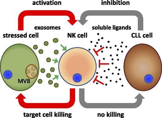
The only variation reported so far for NK cells from CLL patients compared with NK cells from healthy donors is a slight but significant down-regulation of NKp30.15 A decreased expression of NCRs on NK cells correlating to poor prognostic factors was also reported.20 A critical role for NKp30 for immune surveillance was already proposed but just recently it was demonstrated in gastrointestinal tumors.46 Inhibitory NKp30 splice variants were identified that affected the prognosis of gastrointestinal sarcoma upon treatment with NK cell-stimulatory KIT tyrosine kinase inhibitors.46 Complementary, we uncover a not yet described role for the NKp30-ligand BAG6 in anti-tumor immunity in CLL. This is in line with a concept suggesting that the lack of activating NK cell ligands (either on target cells or on released exosomes) results in a failure of “induced self-activation” of NK cells. This means that activation of NK cells by extracellular ligands “in trans” is possible, which poses the question of how NK cells may retain their specificity against the damaged (and exosomes releasing) target cells. One possible explanation is that NK cells need a second signal as previously discussed47,48 that is delivered by direct cell-cell contact. The ability of tumor cells to deliver this activation is probably lost during tumor progression, as suggested in the Burkitt’s lymphoma Eμ-myc mouse model.48 Based on our data, we propose that the first signal delivered by damaged or transformed target cells relates to exosomes that present activating ligands such as BAG6. A failure of exosome stimulation results in NK cell anergy, further driven by soluble inhibitory factors in the microenvironment (see Figure 7). This model convincingly explains why CLL cells are resistant to resting NK cells, but are sensitive toward activated NK cells.15 Moreover, it opens up potential therapeutic strategies using exosomes as immunotherapeutic agents to restore NK cell function in CLL patients. In conclusion, our data suggest that exosomal BAG6 represents a crucial component of “induced self-activation” of NK cells that is impaired in CLL contributing to immune escape in this disease.
Model for the role of soluble and exosomal BAG6 in NK cell activation and anti-tumor immunity. Cells release BAG6-exosomes in response to cellular stress to alert the NK cells to the dangerous cells. Activation of NK cells through released exosomes triggers target cell recognition and killing. Failure of NK cell activation in combination with the release of soluble ligands, including BAG6 inhibits NK cell function leading to immune evasion of the tumor cells from NK cell attack. MVB, multivesicular bodies.
Model for the role of soluble and exosomal BAG6 in NK cell activation and anti-tumor immunity. Cells release BAG6-exosomes in response to cellular stress to alert the NK cells to the dangerous cells. Activation of NK cells through released exosomes triggers target cell recognition and killing. Failure of NK cell activation in combination with the release of soluble ligands, including BAG6 inhibits NK cell function leading to immune evasion of the tumor cells from NK cell attack. MVB, multivesicular bodies.
The online version of this article contains a data supplement.
The publication costs of this article were defrayed in part by page charge payment. Therefore, and solely to indicate this fact, this article is hereby marked “advertisement” in accordance with 18 USC section 1734.
Acknowledgment
The authors thank Gisela Schön und Petra Mayer for their technical assistance, the blood donors for their generous contribution, and Prof. Birgit Gathof (University of Cologne) for support.
This study was supported by grants from the Deutsche Forschungsgemeinschaft (SFB832), (TP19) (E.P.v.S.), and (TP17) (M.K.).
Authorship
Contribution: K.S.R. and E.P.v.S. designed research; K.S.R., D.T., A.H., V.R.S., J.K., M.S., M.B., and S.T. performed research; K.S.R., D.T., A.H., J.K., H.P.H., and E.P.v.S. analyzed data; M. Herling, M.K., and M. Hallek contributed reagents; and E.P.v.S., K.S.R., A.H, H.P.H., M. Hallek, and M.S. wrote the paper.
Conflict-of-interest disclosure: The authors declare no competing financial interests.
The current affiliation for D.T. is the Department of Pediatric Hematology and Oncology, University of Bonn, Bonn, Germany.
The current affiliation of V.R.S. is the Division of Monoclonal Antibodies, Center for Drug Evaluation and Research, Food and Drug Administration, Bethesda, MD.
Correspondence: Elke Pogge von Strandmann, University of Cologne, Department I of Internal Medicine, Laboratory for Immunotherapy, Kerpener Str. 62, Cologne 50937, Germany; e-mail: elke.pogge@uk-koeln.de.
References
Author notes
K.S.R. and D.T. contributed equally to this study.

![Figure 2. BAG6-positive exosomes trigger NK cell cytotoxicity. (A) BAG6 was detected on exosomes released by 293T cells either constitutively or in response to cellular stress, including treatment with heat shock (HS, 30 minutes, 42°C and 2 hours recovery), doxorubicine (50 nM), TNF-α (50 ng/mL), and valproate (100 μg/mL). Mean and standard error of the mean (SEM) are indicated (n = 3), the enhanced BAG6 release was significant compared with untreated cells (HS: P = .0158; doxo: P = .0142, TNF-α: 0.0037, valproate: P = .001, unpaired Student t test). (B) Flow cytometry demonstrated binding of a BAG6-specific antibody (3E4) and a CD9-specific antibody to exosomes from wt cells (dotted line, endosomes) and exosomes from BAG6-transfected cells (+BAG6, black line). Gray-filled histogram: isotype control. FACS, fluorescence-activated cell sorter. (C) Western blot (WB) analysis to detect BAG6 and exosome marker (HSP70, CD9, CD81, CD63) in exosomes lysates of wt and BAG6-transfected cells (+BAG6). (D) BAG6-expressing exosomes enhanced the NK cell killing activity in cytotoxicity assays against the NK cell-resistant target cell lines 697 and NALM6 because untreated NK cells were less cytolytic than NK cells treated with exosomes purified from wt 293T cells (+exo). NK cells treated with exosomes from BAG6-transfected 293T cells (+BAG6-exo) demonstrated the highest killing activity. One representative of 3 experiments is shown. Blocking of NKp30 (clone P30-15 and IgG1-isotype control from BioLegend) reduced the target cell killing significantly (mean and SEM [n = 3]); 697: P = .0200 and Nalm6: P = .0123, unpaired Student t test; effector:target ratio, 10:1).](https://ash.silverchair-cdn.com/ash/content_public/journal/blood/121/18/10.1182_blood-2013-01-476606/4/m_3658f2.jpeg?Expires=1769116806&Signature=pQhOK7ZJ-5M9Q3BmEarPVmpaCZTIBicKzqAsWBYQlr4ybfkjPbuDBUQVtuKXy4EjLzSQxELk0JJiBB3LuMfIoI-BvaI82NA8EOHW8QHRwyhDaBdPvRrspILW0AbdqsbmnIsWi3XLdRxX4E7sFo6xN3WBzVvLP877XtxtL9YXS5Ojqry0yiBaAlUSSGFPk0v~-~agx4yZVQXbqBIFFEKoA2DjMM~fmh8XQNr27~mCWAwvi6ze90~HMeXb9iM-R2BoFzTOedu2GuL92AWh53898LKbxzx-~xcuxE84Iu7zg1e8j0j3Y2W1FMRM3u9asAEi0OaJlu6vXWuTwxU1OsT1dg__&Key-Pair-Id=APKAIE5G5CRDK6RD3PGA)
